
Strategic short title:
ROP: The Preventable Blindness Pathway
Author:
Dr. Samer Al-Diri
Ophthalmologist and Health-Systems Governance Researcher
Professional profiles: ORCID iD: 0009-0004-1908-0714 | ResearchGate: Samer Al-Diri | Website: drsameraldiri.com
Publication type:
Evidence-informed narrative and policy analysis addressing clinical care, public health, policy, health-system governance and education.
Publication date:
20 July 2026
Suggested citation:
Al-Diri S. Retinopathy of Prematurity: A Global Health-Systems Framework for Preventing Childhood Blindness through Neonatal Safety, Screening Governance, and Lifelong Vision Protection. DrSamerAlDiri.com. 2026. Available from: https://drsameraldiri.com/retinopathy-of-prematurity-global-health-systems-framework/ |DOI: https://doi.org/10.13140/RG.2.2.23814.74566
Educational disclaimer:
This article is intended for educational, professional, and public-health discussion. It does not replace clinical examination, local screening protocols, neonatal care pathways, specialist ophthalmology assessment, or urgent medical advice for any individual infant.
Copyright and licensing statement: © 2026 Dr. Samer Al-Diri. This work is licensed under the Creative Commons Attribution–NonCommercial–NoDerivatives 4.0 International Licence (CC BY-NC-ND 4.0).
Permissions and correspondence: For permissions beyond the terms of the CC BY-NC-ND 4.0 licence, including translations, adaptations, derivative works or commercial use, please contact the author at sameraldiri@aol.com.
Executive Briefing for Leaders
Retinopathy of Prematurity is a global test of health-system integrity. It is a retinal disease of premature infants, but it also reveals whether neonatal survival is matched by safe oxygen governance, timely screening, treatment access, reliable follow-up, and long-term vision protection. [1, 3, 14, 18, 19]
The recurring points of failure are well recognised. An eligible infant may never enter the screening pathway; the first examination may occur late; uncertain findings may not be escalated; treatment-requiring disease may not receive urgent action; or surveillance may be lost after treatment, discharge or transfer [1, 3, 14, 30, 31, 34]
The leadership standard is therefore simple but demanding: no eligible infant should be invisible, no screening window should be missed, no severe disease should be left without urgent action, no treatment should occur without surveillance, and no discharge should happen without documented retinal status. [1, 3, 14, 30, 31, 34]
Immediate priorities include maintaining a live ROP register, assigning named responsibility, auditing delay and loss to follow-up rather than activity alone, and confirming treatment, referral and transfer arrangements before an urgent case arises. [1, 3, 30, 31, 34] Parents need clear information and practical support, without being expected to carry responsibility for weaknesses in the pathway. [1, 3] Tele-ROP and artificial intelligence should be governed as components of clinical care, not treated as stand-alone solutions. [35, 36, 41, 42, 43, 46]
To make these risks visible and measurable, this article brings together the NICU-to-Retina Safety Chain, the ROP Blindness Prevention Cascade, the Survival–Vision Paradox and Lifelong Vision Debt, while adapting the established Third Delay concept to the ROP pathway. These constructs are intended to support programme design, evaluation, accountability and implementation.
The central conclusion is straightforward: a written guideline protects vision only when it is translated into a functioning pathway. That pathway must reliably identify, examine, document, escalate, treat and follow every eligible infant from neonatal care into long-term vision protection. [30, 31, 33, 34]
Contents
Use the links below to navigate directly to sections, tables, glossary entries, and reference groups within this article.
Executive Briefing for Leaders
Interpretive Boundaries and Scholarly Caution
Executive Summary for Policymakers and Healthcare Leaders
Five priority actions for ROP blindness prevention
Part I — What Retinopathy of Prematurity Is and Why It Matters
1.1 The developing retina and the premature infant
1.2 Why ROP can progress silently
1.4 ROP as a dynamic disease, not a fixed diagnosis
1.5 When ROP becomes vision-threatening
1.6 Why ROP matters beyond the eye
1.7 ROP as a health-system test
Table 1. ROP at a glance: clinical, public-health, and system meaning
Part II — Global Burden and Public Health Significance
2.1 Prematurity as the starting point of the burden
2.2 The changing global pattern of ROP
2.3 Middle-income and resource-constrained settings
2.4 ROP and preventable childhood blindness
2.5 Equity and the geography of risk
2.6 The hidden burden after survival
2.7 Why ROP is a public-health systems priority
2.8 The Survival–Vision Paradox as a leadership warning
2.9 Active surveillance as a burden-estimation lesson
Part III — Screening: The Time-Critical Prevention Pathway
3.1 Screening begins with eligibility recognition
3.2 The first examination must be timed, not guessed
3.3 Screening is not complete after one examination
3.4 Accurate grading is part of screening safety
3.5 Documentation is a clinical safety tool
Table 2. ROP documentation checklist: minimum safety record elements
3.6 Discharge safety: the pathway must not stop at the neonatal door
3.7 Transfer communication: no infant should move without their retinal status
3.8 Parent counselling without parent-blaming
3.10 Screening governance: from appointment to accountability
3.11 Screening burden, workforce sustainability, and safe thresholds
Part IV — Treatment and Follow-up
4.1 Parent-facing treatment summary
4.2 When ROP becomes treatment-requiring
4.6 Laser and anti-VEGF are not simply competing treatments
4.8 Post-treatment surveillance
4.9 Recurrence, reactivation, and retreatment
4.10 Long-term eye care after ROP
4.11 Treatment is a multidisciplinary event
Table 4. Treatment options: laser, anti-VEGF, surgery, follow-up, and key cautions
4.12 Follow-up governance: the treatment pathway is not complete until risk is closed
Part V — Where ROP Programmes Fail: The Delay Chain and the Third Delay
5.1 ROP blindness as a pathway failure
5.2 The three delay domains in ROP blindness prevention
5.3 Screening delay and grading/decision quality are related but distinct
5.4 The Third Delay: after diagnosis but before protection
5.5 Data invisibility and underestimated burden
5.6 Recurrence, retreatment, and the danger of premature reassurance
5.7 Programme response: treat delay as a safety event
Part VI — Building a Safe ROP Screening System
6.1 The NICU-to-Retina Safety Chain
6.2 The ROP Blindness Prevention Cascade
6.3 Governance roles: who owns each part of the pathway?
6.4 The ROP register as the operational backbone
6.5 Scheduling and due-list review
6.6 Escalation pathways for urgent disease
6.7 Discharge and transfer controls
6.8 Parent communication as part of system design
Table 5. ROP programme safety dashboard: domains, indicators, and safety meaning
6.10 Multidisciplinary accountability
6.11 Minimum safe system requirements
6.12 From safe system to mature programme
Table 6. ROP programme maturity model: basic, developing, functional, advanced, and AI-enabled
6.14 From national surveillance to local operational dashboards
Part VII — The Governance of AI in ROP Screening
7.1 Why AI and tele-ROP matter
7.2 Tele-ROP as the foundation for AI-supported screening
7.3 Where AI can support the ROP pathway
7.4 What AI must not be asked to do
7.5 Validation before adoption
7.6 Data quality and imaging governance
7.7 Human oversight and clinical accountability
7.8 Bias, equity, and local performance
7.9 AI must be linked to treatment access
7.10 Legal, ethical, and professional responsibility
7.11 Implementation in resource-constrained settings
Table 7. AI and tele-ROP governance: opportunities, risks, safeguards, and accountability
7.12 Minimum governance requirements before AI deployment
7.13 From technology adoption to governed innovation
Part VIII — Comparative Policy Landscape
8.1 From guideline to governance
8.2 United Kingdom: national guidance with separated screening and treatment responsibilities
8.3 United States: professional society policy and programme responsibility
8.4 Australasia: guideline-based care with emphasis on local protocol and team responsibility
8.5 India: broader eligibility, public-health scale, and implementation innovation
8.6 Resource-constrained and middle-income settings: adapting principles, not copying thresholds
8.7 Screening mandates and accountability
8.8 Audit: the difference between policy presence and policy performance
8.9 Treatment access as a policy requirement
8.10 Parent information and public trust
8.11 Comparative lessons for policy design
Table 8. Comparative policy landscape: UK, US, Australasia, India, and resource-constrained settings
8.12 Policy as a pathway, not a document
Part IX — Equity, Ethics, and Lifelong Impact
9.1 ROP blindness as an equity failure
9.2 The moral weight of early-life prevention
9.4 Beyond anatomical success: functional vision and participation
9.5 Education and school readiness
9.6 Family burden and parental wellbeing
9.7 The social meaning of preventable childhood blindness
9.8 Ethical care without parent-blaming
9.9 Equity for rural, remote, and under-resourced neonatal units
9.10 Disability rights and inclusive support
9.11 Productivity, economic participation, and social investment
9.12 Equity in AI and digital ROP systems
9.13 From neonatal safety to life-course justice
9.14 The ethical test for health-system leaders
3. Minimum safe-system requirements
Table 9. Policy brief minimum safe-system requirements: essential ROP safety architecture
4. Priority actions for neonatal units
5. Priority actions for ophthalmology departments
6. Priority actions for hospital executives
7. Priority actions for ministries of health and regional networks
8. Priority actions for NGOs and child-eye-health programmes
9. Priority actions for AI, tele-ROP, and digital-health programmes
10. The ROP Blindness Prevention Cascade for audit
Table 10. ROP Blindness Prevention Cascade audit dashboard: minimum programme indicators
11. Red flags requiring urgent programme review
12. Implementation priorities: first 100 days
14. Longer-term system priorities
B. Professional and Leadership FAQs
Definitive Glossary of ROP and Neonatal Safety Terminology
A1. Prematurity, retinal development, and core ROP terms
Table 11. Glossary A1: prematurity, retinal development, and core ROP terms
A2. ROP classification, activity, and severity terms
Table 12. Glossary A2: ROP classification, activity, and severity terms
B. Treatment, recurrence, and long-term vision protection
Table 13. Glossary B: treatment, recurrence, and long-term vision protection
C1. Screening eligibility, follow-up, and register terminology
Table 14. Glossary C1: screening eligibility, follow-up, and register terminology
C2. Governance, transfer, audit, and oxygen-safety terminology
Table 15. Glossary C2: governance, transfer, audit, and oxygen-safety terminology
D. Original frameworks used in this article
Table 16. Glossary D: original frameworks used in this article
E. Digital health, tele-ROP, and AI governance terms
Table 17. Glossary E: digital health, tele-ROP, and AI governance terms
F. Public-health and equity terms
Table 18. Glossary F: public-health and equity terms
Methods and evidence statement
AI and digital-health disclaimer
Open-access and reuse statement
Table 19. Abbreviations used in the article
Core guidelines, classification standards and treatment trials
UK surveillance, treatment patterns and service-delivery evidence
Oxygen governance, neonatal risk and global burden
Anti-VEGF recurrence, systemic safety and follow-up governance
Patient safety, delay frameworks and health-system governance
Tele-ROP, AI-enabled screening and digital governance
Long-term visual, refractive and developmental outcomes
Global vision, child development and rights frameworks
Evidence and methodology note
This article draws on international ROP classification standards, national screening and treatment guidance, landmark clinical trials, population surveillance studies, oxygen-target evidence, anti-VEGF safety literature, tele-ROP and AI validation studies, patient-safety science, public-health delay frameworks, and global child-health and disability-rights sources.
This article employs a critical interpretive synthesis of the international ROP literature to develop a systems-level governance framework. Where existing terminology did not adequately capture important health-system relationships, original conceptual constructs were developed to strengthen analytical clarity, programme evaluation, and translation into practice.
Within this framework, the NICU-to-Retina Safety Chain, the ROP Blindness Prevention Cascade, the Survival–Vision Paradox, and Lifelong Vision Debt are author-developed synthesis constructs. The Third Delay in ROP Blindness is an author-developed adaptation of the established Three Delays model from maternal-mortality analysis to the ROP pathway. [32] These constructs are intended to support governance, audit, accountability and translation into practice; they have not been externally validated as composite clinical or programme tools and do not replace local clinical protocols, national eligibility criteria, specialist judgement or urgent treatment pathways.
Interpretive Boundaries and Scholarly Caution
This article proposes a health-systems framework for understanding and preventing avoidable blindness from Retinopathy of Prematurity. The frameworks introduced here – including the NICU-to-Retina Safety Chain, the ROP Blindness Prevention Cascade, the Third Delay in ROP Blindness, the Survival–Vision Paradox, and Lifelong Vision Debt – are intended to help clinicians, neonatal teams, managers, planners, and policymakers see where risk accumulates and where accountability must be designed. They should be read as conceptual and operational tools, not as replacements for national screening guidelines, local clinical protocols, specialist ophthalmic judgement, neonatal safety standards, or formal treatment criteria.
ROP prevention remains context-specific. Screening eligibility, examination timing, treatment thresholds, laser and anti-VEGF decisions, post-treatment surveillance, and long-term follow-up must be interpreted in the clinical context of the individual infant and within recognised professional guidance. The same caution applies to tele-ROP and AI-supported screening: technology may strengthen a pathway, but it does not remove the need for clear responsibility, treatment access, parent communication, data governance, and safety oversight.
For that reason, the article is deliberately written in the language of safety and learning rather than blame. Its purpose is not to assign failure to parents, clinicians, neonatal units, ophthalmology teams, hospitals, or policymakers. Its purpose is to make preventable points of harm visible, so that systems can be designed to protect infants more reliably. The central argument is therefore clinical, operational, and ethical: preventing ROP blindness requires both knowledge of the disease and a dependable pathway capable of converting neonatal survival into lifelong vision protection.
A final critical balance is important. A systems approach does not dilute individual clinical responsibility; it makes responsible clinical practice more reliable. Skilled examination, sound judgement, accurate documentation, urgent escalation, treatment competence, and parent communication remain essential. The purpose of governance is to ensure that these professional responsibilities are supported by registers, scheduling systems, escalation routes, treatment access, audit, and institutional accountability, rather than being left to memory, goodwill, or chance. [30, 31, 33, 34]
Executive Abstract
Retinopathy of Prematurity (ROP) is often introduced as a retinal disease of premature infants. That description is clinically correct, but incomplete. ROP is also a neonatal safety issue, a screening-governance challenge, a preventable childhood-blindness pathway, and a life-course public-health priority. [1, 3, 14, 18, 19]
The condition arises in the setting of premature retinal vascular development, where the immature retina is vulnerable to disordered vascular growth. International classification systems describe ROP using anatomical and severity features such as retinal zone, disease stage, extent, plus disease, and aggressive ROP. Yet the most important question for health systems is not only whether ROP can be identified. It is whether every eligible infant is recognised, screened at the right time, graded accurately, treated within the therapeutic window when indicated, followed after treatment or regression, and protected from avoidable long-term visual disability.
Globally, the relevance of ROP is increasing because neonatal survival has improved in many settings. This is a major achievement. However, survival must be matched by safe oxygen governance, structured screening, reliable referral, timely treatment, and long-term follow-up. Without these safeguards, a child who survives prematurity may still face avoidable visual impairment or blindness.
This article reframes ROP blindness as the visible endpoint of a pathway failure rather than as an isolated biological event. The present analysis introduces and defines a health-systems framework built around the NICU-to-Retina Safety Chain, the ROP Blindness Prevention Cascade, the Third Delay in ROP Blindness, the Survival–Vision Paradox, and Lifelong Vision Debt. These frameworks are intended to help clinicians, neonatal teams, hospital leaders, policymakers, and digital-health innovators move beyond awareness alone toward measurable accountability. Together, these original synthesis frameworks give the article its translational purpose: while the clinical ROP literature defines what should be recognised, monitored and treated, this article focuses on how those responsibilities can be delivered reliably, equitably and accountably across neonatal, ophthalmology, policy and digital-health systems.
AI-supported retinal imaging and tele-ROP models may strengthen screening capacity, particularly where specialist access is limited. However, technology cannot replace clinical responsibility, treatment access, parent communication, data governance, or system accountability. In ROP, innovation is only safe when it is embedded within a reliable pathway from neonatal admission to lifelong vision protection.
Preventing ROP blindness is therefore not a narrow ophthalmic task. It is a test of whether modern health systems can convert neonatal survival into protected childhood development, educational opportunity, family wellbeing, and lifelong visual function.
Executive Summary for Policymakers and Healthcare Leaders
Retinopathy of Prematurity (ROP) is a preventable cause of childhood blindness affecting premature infants. It is not only an ophthalmic diagnosis, but a test of whether neonatal survival is matched by safe screening, timely treatment, and reliable long-term follow-up. [1, 3, 14, 18, 19]
Prevention must nevertheless be described precisely. Some infants develop severe disease or visual loss despite timely, appropriate and well-governed care. The preventable focus of this article concerns avoidable harm arising from missed eligibility, delayed or incomplete screening, uncertain grading, treatment delay, inadequate discharge or transfer arrangements, and loss to follow-up. It does not imply that every adverse retinal outcome can be eliminated. [1, 3, 14, 30, 31, 34]
The global importance of ROP has increased as more premature infants survive. This progress should be recognised as a major achievement of neonatal care. However, survival alone is not the final endpoint of a safe neonatal system. For premature infants at risk of ROP, survival must be connected to vision protection through oxygen governance, eligibility recognition, scheduled retinal screening, accurate disease classification, timely treatment where indicated, and continued surveillance after discharge.
ROP prevention requires a coordinated pathway. No single professional group can carry the responsibility alone. Neonatal teams, ophthalmologists, nurses, hospital managers, ministries of health, imaging services, referral networks, and parents all form part of the safety chain. A premature infant may lose the opportunity for sight-saving care not because ROP is untreatable, but because the pathway fails at one or more predictable points: the infant is not recognised as eligible, the examination is missed or delayed, the disease is not graded accurately, treatment is not delivered within the required window, or follow-up is lost after discharge or transfer.
For health-system leaders, the central message is clear: Avoidable ROP blindness should be investigated as a potential pathway failure, while recognising that not every adverse retinal outcome is preventable. This does not mean blaming individual clinicians, neonatal teams, or parents. It means designing systems in which the right infant is identified, examined, documented, escalated, treated, and followed with enough reliability that preventable blindness becomes increasingly rare.
Five priority actions for ROP blindness prevention
1. Make ROP screening a formal neonatal safety pathway, not an optional referral.
Every neonatal unit caring for premature infants should have a written ROP screening protocol, clear eligibility criteria, a named responsible team, and a system for ensuring that no eligible infant is discharged, transferred, or lost without a documented screening plan. [1, 3, 4]
2. Build an accountable ROP register from eligibility to safe completion of ROP surveillance. A safe ROP programme requires a live register that tracks eligibility, first examination timing, repeat examination intervals, disease classification, treatment decisions, post-treatment surveillance, and final outcome. The register should not stop at NICU discharge, because many failures occur during transfer, outpatient follow-up, or the transition between neonatal and ophthalmology services.
3. Audit delay, not only activity.
Counting the number of infants screened is not enough. Programmes should measure whether eligible infants were identified on time, whether screening occurred within the required window, whether findings were documented clearly, whether treatment-requiring disease was escalated urgently, and whether follow-up appointments were completed. Missed examinations, late examinations, uncertain grading, delayed treatment, failed transfers, and follow-up loss should be reviewed as safety events.
4. Strengthen the workforce, equipment, and referral network before crisis occurs.
ROP prevention depends on trained examiners, competent graders, reliable imaging where used, safe treatment access, neonatal-ophthalmology communication, and agreed escalation routes for infants who require urgent treatment. In regions without enough specialist ophthalmologists, tele-ROP and wide-field imaging may extend access, but only when embedded within clinical governance, treatment availability, quality assurance, and clear responsibility for action.
5. Treat AI as a governed support tool, not a replacement for accountability.
AI-supported ROP screening may improve capacity, consistency, and risk detection, especially in underserved settings. However, AI cannot compensate for absent screening pathways, weak referral systems, poor image quality, delayed treatment, or unclear professional responsibility. AI in ROP should be clinically validated, locally monitored, ethically governed, and connected to a pathway where positive or uncertain findings lead to timely human review and treatment access.
These five actions are interdependent. A register without a protocol has nothing reliable to track. A protocol without trained staff and equipment cannot be implemented safely. An audit system without a live register has no dependable data. AI without a functioning clinical pathway may create risk rather than reduce it. Leaders should therefore begin with one honest question: does every premature infant in our care have a named professional responsible for ensuring that ROP screening, documentation, escalation, and follow-up are completed? If the answer is no, the first step is to close that accountability gap before adding complexity.
ROP prevention is therefore a leadership responsibility as much as a clinical responsibility. A safe programme does not depend on awareness alone. It depends on governance, measurement, communication, escalation, and accountability across the full NICU-to-retina pathway.
Preventing ROP blindness protects more than the retina. It protects early childhood development, educational opportunity, family wellbeing, and the life-course potential of infants who have already survived a vulnerable beginning.
Plain-Language Parent Summary
If your baby was born very early, with a very low birth weight, or had a difficult neonatal course, the neonatal team may recommend eye screening for a condition called Retinopathy of Prematurity, often shortened to ROP. The exact screening criteria vary between countries, neonatal units, and local guidelines, but they usually consider gestational age, birth weight, and the baby’s overall medical risk. This can sound worrying, especially when your baby is already receiving care in a neonatal unit. The most important message is this: ROP screening is not done because something has definitely gone wrong. It is done because premature babies need careful protection while their eyes are still developing. [1, 3, 4]
The retina is the light-sensitive layer at the back of the eye. In babies born at full term, the blood vessels of the retina have usually had enough time to grow before birth. In premature babies, this development may still be incomplete. After birth, the immature retina continues to grow, but in some babies the growth of retinal blood vessels can become abnormal. This is what doctors call Retinopathy of Prematurity.
Many babies who are screened for ROP do not develop severe disease and do not need treatment. Some babies develop mild ROP that improves on its own with careful monitoring. A smaller number develop more serious ROP that may need treatment to protect sight. Screening matters because the condition can change over time, and the safest approach is to detect concerning changes early, before they threaten vision.
ROP is not the fault of parents. It is not caused by anything a mother or father did or failed to do. It is linked to prematurity, early retinal development, neonatal illness, oxygen exposure, and the complex medical needs of very small or very early babies. Oxygen can be lifesaving in neonatal care, but it also needs careful monitoring because the immature retina is sensitive during this stage of development.
The purpose of ROP screening is to check whether the blood vessels in the retina are developing safely. The eye examination is usually performed by a trained ophthalmologist or appropriately trained specialist team. Eye drops may be used to make the pupils larger so that the retina can be examined properly. Some babies are examined directly, while others may have retinal photographs taken using specialised wide-field imaging equipment. The examination can be uncomfortable for a short time, but it is performed to protect the baby’s long-term vision.
Timing is extremely important. ROP does not always appear immediately after birth, and the highest-risk period may occur while the baby is still in the neonatal unit or after transfer or discharge. This is why parents may be given repeat appointments. These appointments are not routine in the ordinary sense; they are part of a time-sensitive safety pathway. Missing or delaying follow-up can mean that important changes are not detected at the right time. In a small number of babies, missed or delayed detection can allow the disease to become harder to treat and may cause lasting damage to vision.
If treatment is needed, the ophthalmology team will explain the findings and the recommended options. Treatment may include laser treatment, a carefully administered injection of medicine into the eye under specialist conditions, or, in more advanced cases, surgery. Not every baby with ROP needs treatment. The decision depends on the severity, location, and behaviour of the disease, and on the specialist assessment of whether the baby’s sight is at risk. If an anti-VEGF injection is used, follow-up may need to continue for longer because ROP can reactivate later.
Even after ROP improves or treatment has been completed, follow-up may still be important. Babies born prematurely can have a higher risk of later vision problems such as refractive error, the need for glasses, squint, amblyopia, or other developmental visual concerns. Long-term eye care is therefore not only about preventing blindness. It is also about supporting the child’s visual development, learning, confidence, and quality of life.
Parents are an essential part of the safety pathway, but they should not be made to feel responsible for carrying the system alone. Before leaving the neonatal unit, parents should feel able to ask clear questions: Does my baby need ROP screening? Has the first eye examination been done? When is the next appointment? Where will it take place? Who should I contact if the appointment letter does not arrive? What should I do if my baby is transferred to another hospital or discharged before screening is complete?
The safest ROP pathway is one in which parents, neonatal teams, ophthalmology teams, and hospital systems all work together. A baby should not depend on memory, chance, or parental persistence alone to receive sight-saving screening. There should be a clear plan, a documented appointment, a responsible team, and a reliable follow-up system.
For families, the message should be realistic but reassuring. ROP can be serious, but it is also one of the clearest examples of a condition where timely screening, careful monitoring, and appropriate treatment can protect vision. The goal is not to frighten parents. The goal is to make sure that every premature baby who needs eye screening is identified, examined, followed, and treated at the right time.
Part I — What Retinopathy of Prematurity Is and Why It Matters
Key Concept:
ROP is not a passive outcome of prematurity. It is an active pathological process at the interface of oxygen exposure, growth factors, neonatal physiology, systemic illness, and the immature retinal vasculature. [16, 17, 18]
Retinopathy of Prematurity, or ROP, is a disease of abnormal retinal vascular development that affects premature infants. It occurs because the retina, particularly its blood-vessel network, may still be immature at the time of birth. In a full-term pregnancy, retinal vascularisation usually has more time to progress before delivery. In a premature infant, this developmental process may be interrupted and then forced to continue outside the protective environment of the womb, during a period when the baby may also require oxygen support and respiratory care, infection treatment, blood transfusion, nutritional support, and the broader intensive care necessary for survival.
ROP should therefore be understood as more than a disease label. It is a clinical condition, but it is also a marker of neonatal vulnerability. It sits at the meeting point of premature birth, retinal development, neonatal physiology, oxygen governance, specialist screening, timely treatment, and long-term childhood vision protection. This is why ROP belongs not only in ophthalmology clinics, but also in neonatal safety discussions, hospital governance systems, public-health planning, and health-service design.
1.1 The developing retina and the premature infant
The retina is the light-sensitive tissue at the back of the eye. It receives visual information and begins the process by which light is converted into signals that the brain can interpret. For this tissue to function properly, it requires an organised blood-vessel supply. During fetal development, retinal blood vessels grow gradually across the retina. Premature birth can interrupt this normal sequence before vascularisation is complete. [18]
After premature birth, retinal vascular development continues in an extrauterine environment shaped by the infant’s immaturity, illness and necessary neonatal care. Oxygen and respiratory support may be lifesaving, but the developing retina is sensitive to oxygen exposure and to changes in the growth factors that regulate vascular development. ROP can arise when normal vascularisation is disrupted and abnormal vessels subsequently develop at the junction between vascularised and avascular retina.
This is the biological basis of ROP. It is not a simple matter of “too much oxygen” or one isolated neonatal event. The condition is influenced by prematurity, low birth weight, incomplete retinal vascularisation, oxygen exposure and fluctuation, systemic illness, inflammation, nutrition, and the wider stability of neonatal care. Reducing ROP risk therefore requires good neonatal practice, but preventing ROP blindness requires something more: a reliable screening and treatment pathway.
1.2 Why ROP can progress silently
One of the most important features of ROP is that it may develop without visible signs that parents or non-specialist staff can easily detect. A premature baby may appear to be improving clinically while retinal disease is still evolving. The eye may look normal from the outside. The baby may not show obvious signs of visual difficulty during the early neonatal period. Yet inside the eye, abnormal retinal vascular changes may be progressing. [6, 18]
This is why ROP screening cannot depend on outward appearance, parental concern, or waiting for symptoms. It must be based on eligibility criteria, timing, documented examinations, and specialist interpretation. The pathway must identify infants at risk before severe disease is clinically obvious. Once advanced retinal detachment has occurred, the opportunity for straightforward sight-saving prevention may already have been lost.
In this sense, ROP resembles other preventable vision conditions where the window for effective action is time-sensitive. The challenge is not merely to know that ROP exists. The challenge is to find it at the right moment, understand its severity accurately, and act before the disease crosses the threshold into irreversible harm.
1.3 How ROP is classified
ROP is classified using internationally recognised clinical features. These features help ophthalmologists describe where the disease is, how severe it is, how extensive it is, and whether there are signs of vascular activity that increase concern.[6]
The main classification elements include:
Zone — the location of disease within the retina. Zone I is the most posterior retinal zone, centred on the optic disc and encompassing the macular region. ROP in Zone I is particularly concerning because posterior disease may progress rapidly and carries a greater risk of adverse outcomes.
Stage — the structural severity of disease at the border between vascularised and avascular retina. Stage 1 indicates a demarcation line. Stage 2 indicates a ridge. Stage 3 indicates extraretinal fibrovascular proliferation. Stage 4 indicates partial retinal detachment, subclassified by whether the fovea is spared or involved. Stage 5 indicates total retinal detachment.
Extent — how much of the retina is involved, usually described by clock hours or sectors of disease.
Plus disease — abnormal dilation and tortuosity of the posterior retinal vessels, reflecting increased disease activity. Plus disease is a major marker of severity and is central to treatment decisions.
Pre-plus disease — vascular abnormality that is more than normal but not severe enough to meet the definition of plus disease. It indicates increased disease activity and usually warrants closer follow-up.
For non-specialist readers, the distinction is important: pre-plus disease is a warning sign of abnormal vascular activity that may require closer surveillance, whereas plus disease represents a more severe level of posterior vascular change and can shift management toward urgent review or treatment depending on zone, stage, extent and overall disease behaviour.[6, 7]
Aggressive ROP — a severe and rapidly progressive form of ROP, often posterior, with prominent vascular activity and a higher risk of rapid deterioration if not recognised and treated urgently.
These terms are clinically important because ROP is not a single uniform condition. Mild peripheral ROP that is improving is very different from posterior aggressive ROP or treatment-requiring disease. Accurate classification allows the clinical team to decide whether the infant needs continued observation, shorter-interval follow-up, urgent treatment, or referral to a specialist treatment centre.
1.4 ROP as a dynamic disease, not a fixed diagnosis
ROP is dynamic. It can progress, stabilise, regress, reactivate, or recur after treatment. A single examination is therefore not always enough. The timing of repeat examinations depends on the infant’s retinal findings, gestational age, postmenstrual age, disease zone, disease stage, vascular activity, and whether treatment has been given. [6, 7]
This dynamic behaviour is one reason why ROP screening programmes require discipline. The danger is not only that an infant is never screened. The danger may also be that the first examination is done, but the next examination is missed; that findings are documented without a clear follow-up interval; that an infant is transferred without the ROP status being handed over; or that apparent early improvement leads to premature discharge from surveillance.
A safe ROP pathway must therefore track the infant over time. It must ask not only, “Was this baby examined?” but also, “Was the examination performed at the correct time? Were the findings classified accurately? Was the follow-up interval safe? Was the next appointment completed? Was treatment delivered within the required window when indicated? Was post-treatment surveillance maintained?”
1.5 When ROP becomes vision-threatening
Many infants who develop ROP do not require treatment. Mild disease may regress with careful monitoring. However, some infants develop treatment-requiring ROP. This is the point at which the disease has reached a level where the risk of progression is high enough that intervention is recommended to reduce the risk of retinal detachment and permanent visual loss. [2, 7, 8]
The central clinical concern in severe ROP is traction on the developing retina. Abnormal blood vessels and associated fibrovascular tissue can contract and pull on the retina. If this traction progresses, it may lead to partial or total retinal detachment. Advanced retinal detachment in infancy can have profound consequences for visual development and may be difficult to treat successfully.
This is why ROP is one of the most time-critical conditions in paediatric ophthalmology. There is a period during which screening, classification, and treatment can protect vision. If that period is missed, the clinical problem changes from prevention to rescue, and from straightforward treatment to complex management with a less predictable visual outcome.
1.6 Why ROP matters beyond the eye
ROP matters because it can affect the whole life course of a child. Vision is not only a sensory function. It influences early development, bonding, mobility, learning, communication, independence, education, family life, and social participation. A preventable loss of vision in infancy can shape decades of opportunity. [47, 48, 49, 51, 54]
This is the concept of Lifelong Vision Debt. A missed neonatal screening or treatment opportunity may create consequences that continue long after the neonatal admission has ended. The original failure may occur during a narrow clinical window, but its impact can extend into childhood development, school readiness, family burden, social inclusion, employment potential, and long-term quality of life.
For this reason, ROP cannot be approached only as an acute retinal disease. It should be seen as an early-life public-health priority. Preventing ROP blindness protects more than anatomical sight. It protects developmental potential at the beginning of life.
1.7 ROP as a health-system test
A well-functioning ROP programme requires more than clinical knowledge. It requires a system capable of identifying eligible infants, scheduling examinations, documenting findings, communicating across teams, escalating urgent disease, delivering treatment, supporting parents, and maintaining follow-up across discharge or transfer. [1, 3, 4, 14, 35, 36]
This makes ROP a powerful test of health-system reliability. The disease itself is biological, but the prevention of blindness depends on organisation. A baby does not benefit from the existence of guidelines unless those guidelines are converted into local practice. A neonatal unit does not become safe for ROP prevention simply because screening is theoretically available. It becomes safe when every eligible infant is reliably captured by the pathway.
The most serious ROP failures are rarely caused by absence of knowledge alone. They often arise from gaps between knowledge and execution: unclear responsibility, incomplete registers, delayed examinations, uncertain grading, poor handover, lack of treatment access, equipment gaps, weak referral pathways, and follow-up loss. These are not inevitable features of prematurity. They are design problems in the safety pathway.
Patient-safety science gives this argument its discipline. The point is not that individual staff fail to care. It is that complex clinical work needs systems that anticipate human limitation, handover loss, unclear ownership, equipment gaps and latent risk. In ROP, the vulnerable infant moves across neonatal, ophthalmic, imaging, administrative and family interfaces. A reliable programme therefore behaves like a high-reliability safety pathway: it reduces dependence on memory, makes overdue risk visible, and ensures that every abnormal or uncertain finding has a named route to action.
Table 1. ROP at a glance: clinical, public-health, and system meaning
| Dimension | What it means | Why it matters |
| Clinical meaning | ROP is abnormal retinal vascular development in premature infants. | It can progress from mild disease to treatment-requiring disease and, in severe cases, retinal detachment. |
| Neonatal meaning | ROP occurs in infants whose retinal development continues during intensive neonatal care. | Oxygen governance, systemic stability, nutrition, infection control, and neonatal monitoring all influence risk. |
| Screening meaning | ROP may progress silently and requires timed specialist examination. | The baby may look well externally while retinal disease is evolving. |
| Treatment meaning | Some infants require urgent intervention to reduce the risk of permanent visual loss. | The window for effective intervention is narrow; treatment delay in severe or aggressive disease may alter the visual outcome. |
| Follow-up meaning | ROP can regress, progress, recur, or be associated with later visual problems. | Long-term eye care may be needed for refractive error, amblyopia, strabismus, and developmental vision support. |
| Public-health meaning | ROP blindness is often preventable when screening and treatment systems work reliably. | It represents a preventable childhood-blindness pathway, not only an individual clinical diagnosis. |
| Health-system meaning | ROP prevention depends on coordination between neonatal care, ophthalmology, hospital leadership, referral systems, and parents. | Failure at any point in the pathway can convert neonatal survival into lifelong visual disability. |
ROP is therefore best understood as both a retinal disease and a systems challenge. Its biology explains why premature infants are at risk. Its classification explains how clinicians judge severity. Its public-health significance lies in the fact that blindness can often be prevented when the pathway functions reliably. The next task is to examine this wider public-health significance: why ROP has become more important as neonatal survival improves, and why preventing ROP blindness must be seen as part of a broader global child-health and health-systems agenda.
Part II — Global Burden and Public Health Significance
Key Concept:
The gains achieved through improved neonatal survival can be undermined by preventable visual disability when screening, treatment and follow-up systems do not develop alongside neonatal care.[1, 3, 4, 13, 14, 47]
Retinopathy of Prematurity has become a major global child-eye-health issue because it sits directly inside one of modern medicine’s greatest achievements: the survival of premature infants. As neonatal care improves, more very small and very early babies survive. This progress should be recognised and protected. However, survival alone is not the final measure of success. A safe neonatal system must also protect the child’s long-term development, including vision.
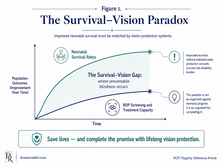
This article introduces the Survival–Vision Paradox in ROP to describe the gap that can emerge when improved neonatal survival is not matched by the systems required to protect vision. The same progress that allows more premature infants to survive can increase the number of babies at risk of ROP if survival is not accompanied by oxygen governance, structured screening, timely treatment, and reliable follow-up. ROP therefore reveals a critical truth about health-system development: saving life and protecting lifelong function must advance together.
2.1 Prematurity as the starting point of the burden
The global burden of ROP begins with the global burden of preterm birth. An estimated 13.4 million babies were born preterm worldwide in 2020. [56] Many survive because of advances in neonatal care, including respiratory support, infection management, nutrition, temperature control, and intensive monitoring. These advances are essential, humane, and life-saving. [18, 19, 20]
Yet prematurity is not a single event that ends at discharge from the neonatal unit. It can create a long arc of developmental vulnerability. The premature infant may leave the neonatal unit alive, but still require follow-up for growth, neurodevelopment, hearing, respiratory health, nutrition, and vision. ROP belongs within this wider post-neonatal responsibility.
For health systems, this means ROP cannot be treated as a small ophthalmology problem at the edge of neonatal care. It is part of the unfinished work of prematurity care. If a country invests in neonatal survival without building the systems required to protect vision, it risks converting a survival success into a preventable disability burden.
2.2 The changing global pattern of ROP
ROP has not affected the world in a uniform way. Historically, different patterns of ROP blindness have appeared as neonatal care evolved. In high-income countries, earlier waves of ROP were associated with the survival of increasingly premature infants and with evolving understanding of oxygen management. Over time, improved neonatal practice, oxygen monitoring, screening guidelines, and treatment access reduced the risk of avoidable blindness, although ROP remains a significant challenge even in advanced systems. [18, 19]
In many middle-income countries, the pattern has been different. Neonatal services have expanded, and more premature infants are surviving, but screening systems, trained personnel, oxygen governance, equipment, referral pathways, and treatment access have not always developed at the same pace. This mismatch has contributed to what is often described as a newer global wave of ROP blindness.
This distinction matters because ROP risk is not determined by national income alone. It is determined by the relationship between neonatal survival, quality of neonatal care, screening coverage, treatment capacity, and follow-up reliability. A system may have advanced neonatal units but weak ophthalmic linkage. Another may have capable ophthalmologists but insufficient neonatal tracking. A third may have screening guidelines on paper but poor implementation across districts or private facilities. ROP becomes dangerous when the pathway is incomplete.
The evidence on oxygen-target trials strengthens, rather than simplifies, this upstream prevention message. Oxygen is lifesaving, and ROP prevention should never be reduced to the crude instruction to restrict oxygen. Large neonatal trials showed the clinical tension clearly: lower oxygen saturation targets may reduce severe ROP among survivors, but may also increase mortality risk. [16, 17] The governance lesson is therefore not “less oxygen”; it is safer oxygen governance – monitored saturation targets, avoidance of wide fluctuations, functioning pulse oximetry, trained neonatal practice, alarm discipline, documentation, and audit of oxygen delivery.
2.3 Middle-income and resource-constrained settings
ROP is particularly important in middle-income and resource-constrained settings because these health systems may be in transition. Neonatal survival may improve rapidly, but the supporting safety infrastructure may lag behind. This creates a vulnerable period in which more premature infants survive, but not all are reliably identified, screened, referred, treated, or followed. [18, 19, 20, 23, 24, 25]
The problem is rarely a single missing component. It may include several overlapping constraints: limited numbers of trained ROP examiners, shortage of wide-field retinal imaging, inconsistent oxygen monitoring, lack of neonatal-ophthalmology coordination, weak referral systems, inadequate data collection, uneven coverage between urban and rural areas, and financial or transport barriers for families. In some places, a baby may be saved in a neonatal unit but then lose the opportunity for sight-saving care because the eye pathway has not been built with the same seriousness as the survival pathway.
This is why ROP is not only a disease of prematurity. It is a disease of system transition. It becomes most dangerous when neonatal capacity grows faster than screening governance. In such settings, the public-health priority is not only to increase awareness, but to build practical systems: eligibility registers, named responsibility, screening schedules, referral agreements, treatment access, parent communication, and audit of missed or delayed care.
2.4 ROP and preventable childhood blindness
Childhood blindness has consequences that extend across an entire lifetime. Vision loss in infancy affects early development, communication, mobility, education, independence, family life, and social participation. The human cost is carried by the child, the family, the education system, and society. [18, 19, 20, 21, 22, 23]
ROP is especially important because severe visual loss may often be preventable if the pathway works. This makes ROP different from conditions where prevention or treatment may be limited by biology alone. In ROP, the critical failure is often not that the disease is unknowable or untreatable. The failure is that the infant does not reach the right examination, at the right time, with the right interpretation, followed by the right treatment and surveillance.
Preventing ROP blindness therefore has a high public-health value. It protects vision at the start of life. It reduces the burden of avoidable disability. It supports child development and school readiness. It also reflects the quality of neonatal and paediatric systems, because a child’s visual outcome depends on coordination across several services rather than on one isolated clinical encounter.
2.5 Equity and the geography of risk
ROP is also an equity issue. The risk of avoidable blindness is not distributed evenly. It is shaped by where a baby is born, the quality of neonatal care available, whether oxygen is monitored safely, whether screening is mandatory or optional, whether trained examiners are accessible, whether treatment can be delivered quickly, whether parents can attend follow-up appointments, and whether transfer between facilities is managed safely. [19, 20, 23, 24, 36, 51]
A premature baby in a well-resourced neonatal network with a functioning ROP register, clear screening protocol, trained examiners, treatment access, and reliable follow-up has a very different risk pathway from a baby born in a fragmented system where responsibility is unclear. The biology may be similar. The pathway is not.
This is why ROP blindness should be understood as a preventable equity failure when avoidable pathway gaps expose some infants to a risk that others are protected from. Equity in ROP does not mean offering the same written guideline to every facility. It means ensuring that every eligible infant has a realistic pathway to screening, treatment, and follow-up, regardless of geography, hospital type, income, parental education, or transfer history.
2.6 The hidden burden after survival
The burden of ROP is often underestimated because the most visible outcome, blindness, represents only the severe end of the spectrum. Many children who do not become blind may still require long-term eye care. Premature infants and children with a history of ROP may be at higher risk of refractive error, amblyopia, strabismus, reduced visual function, or other developmental visual concerns. [13, 47, 48, 49, 50]
This matters because public-health planning must look beyond the emergency of treatment-requiring ROP. A safe programme must also consider long-term visual development. The goal is not only to avoid retinal detachment. It is to support the child’s best possible functional vision over time.
The concept of Lifelong Vision Debt is important here. A missed or delayed opportunity in the neonatal period may create consequences that continue into childhood, education, social participation, and adulthood. Even when blindness is avoided, incomplete follow-up may leave children with uncorrected or unmanaged visual needs that affect learning and development.
Long-term outcome evidence also supports this wider framing. Children born very prematurely, and children with a history of ROP or ROP treatment, may carry risks that extend beyond the acute retinal episode: myopia or high myopia, anisometropia, amblyopia, strabismus, reduced visual function and developmental vulnerability. For leaders and service planners, this means that the ROP programme should not be judged only by whether retinal detachment was avoided. It should also ask whether the child entered a pathway capable of protecting functional vision, learning and development over time.
2.7 Why ROP is a public-health systems priority
ROP is a public-health priority because it brings together four features that demand organised action.
First, the population at risk can be defined. Premature infants can be identified using gestational age, birth weight, clinical risk, and local eligibility criteria. [1, 3, 4]
Second, the condition has a time-sensitive natural history. There is a period during which screening and treatment can prevent severe visual loss.
Third, the intervention pathway is known. Screening, classification, follow-up, treatment, and long-term monitoring are established components of care.
Fourth, the consequences of failure are lifelong. When ROP blindness occurs, the effect is not limited to the neonatal admission or the eye department. It can shape development, education, family life, social participation, and economic opportunity.
These features make ROP a clear test of whether a health system can convert knowledge into protection. Awareness is necessary, but insufficient. Guidelines are necessary, but insufficient. Equipment is necessary, but insufficient. What matters is whether the entire pathway works reliably for every eligible infant.
ROP therefore belongs in national child-health strategies, neonatal quality programmes, hospital safety systems, eye-health plans, digital-health investment, and health-equity agendas. It should not sit at the margins of policy as a rare specialist disease. It should be recognised as a preventable childhood-blindness pathway that reflects the maturity, coordination, and accountability of the health system.
2.8 The Survival–Vision Paradox as a leadership warning
The Survival–Vision Paradox is an author-developed conceptual framing introduced in this article. It does not suggest that improved neonatal survival itself causes visual loss. Rather, it describes a mismatch that can arise when gains in neonatal survival increase the number of infants requiring ROP surveillance, while oxygen governance, screening, treatment and follow-up capacity do not expand accordingly. It is not an argument against neonatal progress; it is an argument for completing that progress. [18, 19, 20, 24, 25]
For policymakers and healthcare leaders, the implication is clear. Whenever neonatal services expand, vision-protection capacity should be assessed and strengthened in parallel. Where ROP pathways remain incomplete, more surviving infants may experience delayed detection or treatment and therefore remain at risk of potentially avoidable visual loss. [19, 20, 24, 25] Such harm is not inevitable; its likelihood can be reduced through context-appropriate eligibility criteria, reliable screening, timely treatment and continuity of follow-up. [1, 3, 4, 23]
Neonatal success should therefore be assessed not only by survival to discharge but also by whether longer-term visual and developmental needs are recognised and supported. ROP prevention is an integral component of comprehensive neonatal and follow-up care, not a peripheral ophthalmic add-on. [48, 49, 51, 54, 55]
2.9 Active surveillance as a burden-estimation lesson
UK active-surveillance evidence offers an important policy lesson for health systems. Adams, Bunce, Xing, Butler, Long, Reddy and Dahlmann-Noor conducted nationwide population-based case ascertainment through the British Ophthalmic Surveillance Unit and a national collaborative ROP special interest group. They identified 327 infants treated or referred for ROP treatment during a 12-month period and estimated a treatment incidence of 4.0% using an estimated denominator of 8,112 liveborn infants weighing less than 1500 g—approximately 2.5 times the previously estimated incidence. The authors nevertheless acknowledged two limitations to this estimate: practitioner notification could miss cases, and denominator data for part of the United Kingdom were extrapolated. [12]
The implication is not that every country should reproduce the UK surveillance model. The transferable lesson is that service planning requires sufficiently reliable case ascertainment. If ROP treatment demand is underestimated, workforce, equipment, referral, treatment and follow-up capacity may be planned below actual need. Active surveillance, reliable registers and outcome audits are therefore safety tools that can help leaders align neonatal survival with vision-protection capacity. [12, 13, 15, 25]
The next section turns from burden to prevention. If Part II explains why ROP matters globally, Part III explains how screening becomes the time-critical pathway through which preventable blindness is avoided.
Part III — Screening: The Time-Critical Prevention Pathway
Key Concept:
For eligible infants, ROP screening is a time-critical neonatal safety pathway, not an optional ophthalmic appointment.
Screening is the critical link between recognising ROP risk and detecting sight-threatening disease in time for appropriate surveillance or treatment. Because sight-threatening ROP must be detected through retinal screening and aggressive ROP can progress rapidly, prevention of avoidable ROP-related visual loss cannot rely on symptoms, parental concern or informal referral. [1, 3, 4]
A safe programme therefore requires a structured pathway that identifies eligible infants, schedules examinations according to the applicable guideline, documents and classifies retinal findings, determines repeat-examination intervals from those findings, urgently escalates disease meeting referral or treatment criteria, and secures continuity when discharge or transfer occurs before screening is complete. ROP screening is not a single visit by the eye team; it is a coordinated process linking neonatal and ophthalmic care until acute ROP surveillance can be safely concluded under the applicable guideline, with longer-term paediatric ophthalmic follow-up arranged where indicated. [1, 3, 4]
This pathway should not depend on chance, memory, parental persistence or individual goodwill. Eligibility should trigger entry into a screening record or tracking system and calculation and scheduling of the first examination date. Each examination should generate documented findings and a clear disposition: a follow-up interval and scheduled appointment when further screening is required; timely escalation when referral or treatment criteria are met; or documented conclusion of acute screening when the applicable stopping criteria are satisfied. If discharge or transfer occurs before screening is complete, the handover should document retinal status, the next examination date and the service responsible for ongoing follow-up; before the infant leaves, either the outpatient appointment or the receiving unit’s arrangements for continued screening should be confirmed. [1, 3, 4, 30, 31, 33]
3.1 Screening begins with eligibility recognition
The first step in ROP prevention is identifying which infants require screening. Eligibility criteria vary between countries and health systems, but they usually include gestational age, birth weight, and additional clinical risk factors. Many guidelines focus on very premature infants and very low birth weight infants, with local consideration for larger or more mature babies who had an unstable neonatal course or other risk factors. [1, 3, 4]
Eligibility recognition is not a minor administrative task. It is the first safety gate in the ROP pathway. If the infant is not recognised as eligible, nothing else can happen reliably. The examination is not scheduled. The ophthalmology team may not be informed. Parents may not be counselled. Transfer or discharge may occur without a screening plan. A preventable pathway failure can therefore begin before the first eye examination is ever due. [1, 4, 30, 31]
A safe neonatal unit should treat ROP eligibility as a structured checklist item. The question should be asked at admission, repeated during the neonatal stay, reviewed before transfer, and confirmed before discharge. The infant’s gestational age, birth weight and relevant clinical risk factors should be assessed against the applicable screening criteria, with confirmed eligibility triggering entry into the screening pathway and calculation of the first examination date. [1, 3, 4]
In mature systems, eligibility recognition should not depend on one person remembering the guideline. It should be built into neonatal admission documentation, electronic medical records, ward rounds, discharge planning, and multidisciplinary safety checks. Where electronic systems are not available, a paper register and clearly assigned responsibility can still support a safe pathway if used consistently. [1, 4, 30, 31, 33]
3.2 The first examination must be timed, not guessed
ROP screening is time-sensitive because the onset of serious ROP is related to the infant’s developmental age, usually expressed as postmenstrual age. Postmenstrual age combines gestational age at birth with chronological age after birth. This matters because the most premature infants may take longer after birth to reach the period when severe ROP is likely to appear, while more mature at-risk infants may require screening at a different chronological time. [1, 3, 4]
This is why the first examination should not be chosen casually. It should be calculated using the relevant national or local guideline. The aim is to examine the retina early enough to detect sight-threatening disease before it becomes destructive, but not so early that repeated examinations are performed unnecessarily before meaningful retinal findings can be assessed.
A useful principle is that the first examination should be a scheduled safety event, not a vague intention. It should have a date, a responsible team, and a documented plan. The neonatal team should know when the examination is due. The eye team should know which babies are due. Parents should understand why the examination is being arranged. If the baby is transferred before the first examination, the receiving team should receive the screening status and the exact timing requirement.
A missed first examination is not merely a missed appointment. It may be the beginning of a preventable delay chain. If the first examination is late, every subsequent decision may also become late: grading, follow-up interval, treatment referral, consent, treatment delivery, and post-treatment surveillance. The screening clock therefore begins before the ophthalmologist enters the neonatal unit.
3.3 Screening is not complete after one examination
A single normal or mild early examination does not always mean the infant is safe from later ROP. The retina may still be immature, and ROP can emerge later, progress between examinations, or regress over time. [1, 3, 4] After treatment, disease can recur or reactivate, particularly after anti-VEGF therapy, so surveillance must continue for as long as clinically indicated. [26, 28, 29] For many infants, the first screening examination is the beginning of surveillance, not the end of it.
Follow-up intervals should be determined by retinal findings, disease zone, disease stage, vascular activity, postmenstrual age, whether the disease is improving or worsening, and whether treatment has been given. Infants with more posterior disease, plus disease, aggressive features, uncertain findings, or incomplete retinal vascularisation in Zone I or posterior Zone II may require shorter follow-up intervals. Infants with clearly regressing low-risk findings may be followed at longer intervals, according to specialist judgement and the applicable guideline. [1, 3, 4]
The key governance point is simple: every examination must generate a clear next step. The record should not only state what was seen. It should state what must happen next, when it must happen, and who is responsible for ensuring that it happens. A screening note without a follow-up plan is an incomplete safety document.
This is where many programmes fail. The first examination may be completed, but the next appointment is not booked. The disease may be described, but the urgency is unclear. The baby may be transferred, but the receiving unit may not know the retinal status. Parents may be told that follow-up is needed, but not given a confirmed appointment or contact pathway. In ROP, surveillance fails when the next step is assumed rather than secured.
3.4 Accurate grading is part of screening safety
ROP screening is not only about whether an examination occurred. It is also about whether the findings were interpreted accurately and acted upon correctly. A late examination is dangerous, but an inadequately interpreted examination can also be dangerous. The infant may be seen, yet the zone, stage, extent, plus or pre-plus disease, or features of aggressive ROP may not be recognised with sufficient confidence to trigger the correct follow-up or treatment pathway. [1, 3, 4, 6, 7]
This is why screening must be performed or interpreted by appropriately trained and competent clinicians, supported by clear documentation and escalation routes. [1, 3, 4] Each eye should be documented by zone, stage, extent and posterior pole vascular status (normal, pre-plus or plus); aggressive ROP should be noted, and regression, progression or reactivation recorded when applicable. The record should also state whether treatment-requiring disease is present or suspected. [6, 7]
Uncertain grading should not be treated as reassurance. If the examiner is unsure, if the view is poor, if the image quality is inadequate, if the disease appears posterior, or if there are signs of rapid progression, the pathway should move toward escalation rather than delay. Safety depends not on pretending that every examination is definitive, but on recognising when uncertainty itself creates risk. [1, 3, 4, 6, 35]
In image-based or tele-ROP programmes, accurate grading also depends on image quality, trained image acquisition, timely interpretation, clear reporting, and the ability to obtain urgent bedside examination when needed. Imaging may extend access, but it must not create a false sense of safety. A photograph that is not taken, not readable, not interpreted quickly, or not linked to treatment access cannot prevent blindness. [35, 36]
3.5 Documentation is a clinical safety tool
ROP documentation should be treated as a safety instrument, not a clerical afterthought. The record should allow any responsible clinician to understand the infant’s risk status, the examination findings, the urgency, the follow-up plan, and the consequences of delay. [1, 3, 4, 30, 31]
A safe ROP screening record should usually include:
Table 2. ROP documentation checklist: minimum safety record elements
| Documentation element | Why it matters |
| Infant identifiers, gestational age, birth weight, and postmenstrual age | Confirms eligibility and developmental timing. |
| Date and setting of examination | Establishes whether screening occurred within the required window. |
| Method of assessment | Clarifies whether assessment was by direct examination or image-based/tele-ROP review, and whether further specialist review was required. |
| Retinal zone, stage, extent, and vascular activity | Allows severity and progression to be understood. |
| Plus disease, pre-plus disease, or aggressive features | Identifies disease that may require closer follow-up or urgent treatment. |
| Image quality or examination limitations | Makes uncertainty visible rather than hidden. |
| Follow-up interval and exact next step | Converts the finding into an action. |
| Escalation or treatment recommendation | Ensures that treatment-requiring disease is not managed as routine follow-up. |
| Parent communication | Documents that parents were informed of the findings and the importance of follow-up. |
| Transfer or discharge instructions | Protects continuity when the infant leaves the neonatal unit or moves between services. |
Documentation should answer the safety question: if another clinician sees this baby tomorrow, will they know exactly what must happen next? If the answer is no, the documentation is not adequate for a time-critical screening pathway.
3.6 Discharge safety: the pathway must not stop at the neonatal door
Discharge from the neonatal unit is one of the highest-risk points in the ROP pathway. A baby may leave intensive care before retinal vascularisation is complete, before ROP has fully regressed, before treatment follow-up is complete, or before the risk period has safely passed. The infant may appear clinically stable, feeding better, gaining weight, and ready to go home. Yet the retina may still require surveillance. [1, 3, 5, 14]
This creates a common safety gap. Neonatal discharge can be interpreted emotionally and practically as the end of the crisis, while ROP follow-up still remains time-critical. Parents may be exhausted, relieved, anxious, and overwhelmed by multiple appointments. Letters may be delayed. Outpatient appointments may not be booked. The eye team may assume the neonatal team arranged follow-up. The neonatal team may assume the eye service will send an appointment. The family may assume no news means no concern.
A safe discharge process should not allow this ambiguity. Before discharge, the ROP status should be known, documented, and explained. The next appointment should be arranged, not merely recommended. Parents should know where to go, when to attend, who to contact, and why delay matters. The discharge summary should include the ROP findings, the follow-up interval, the responsible service, and any treatment or surveillance concerns.
If the required ROP follow-up appointment cannot be confirmed before discharge, this should trigger prompt escalation to senior neonatal and ophthalmology decision-makers. A documented risk-management plan should identify the responsible service, the arrangements for securing timely follow-up, and the communication route for the family. In a time-critical pathway, “follow up as outpatient” is not sufficient without a confirmed appointment, clear responsibility, and reliable communication arrangements. [1]
3.7 Transfer communication: no infant should move without their retinal status
Neonatal transfer is another major vulnerability. Premature infants may move between hospitals for intensive care, step-down care, specialist treatment, bed capacity, family location, or repatriation. Each transfer creates a risk that ROP status may be lost or misunderstood. [1, 4]
The transfer handover should include whether the infant is eligible for ROP screening, whether screening has started, the date and findings of the last examination, whether treatment has been given, the next examination date, the urgency of follow-up, and the named service responsible for continuing care. This information should travel with the infant as clearly as medication, respiratory status, feeding plan, infection status, or neurodevelopmental concerns.
The receiving team should not have to reconstruct the ROP pathway from incomplete notes. They should be able to see immediately whether the infant is not yet due, due soon, overdue, under surveillance, awaiting treatment, post-treatment, or ready for discharge from screening. Transfer should therefore be treated as an active handover of retinal risk.
In a safe system, ROP transfer communication is not optional. It is part of neonatal safety governance. The infant should not lose sight-saving continuity because they crossed an institutional boundary.
3.8 Parent counselling without parent-blaming
Parents are essential partners in ROP screening, but they should not be made to carry the safety pathway alone. They need clear information because follow-up may continue after discharge, and because missed appointments can have serious consequences. However, the health system should not shift responsibility onto parents in a way that suggests that pathway safety depends only on their vigilance. [1, 3, 4]
Good parent counselling should be calm, practical, and repeated at key points. Parents should understand that ROP screening is precautionary, that many babies do not need treatment, that timing matters, and that repeat appointments are part of protecting vision. They should be told what the examination is for, what the current findings mean, when the next appointment is due, and who to contact if there is any uncertainty.
Parents should also be given written information. Verbal explanation alone may not be enough, especially when families are under stress or when the baby has complex medical needs. Written information should include the reason for screening, the importance of attendance, the next appointment details, warning that delay may be harmful, and contact details for the responsible team.
The aim is not to frighten parents. The aim is to make the pathway visible. Parents should leave the neonatal unit with a clear understanding that ROP follow-up is time-sensitive, but they should also feel that the clinical team has built a reliable system around their baby.
3.9 The missed-window risk
The most dangerous failure in ROP screening is not simply that an appointment is missed. It is that the treatment window may be missed. ROP has a period during which detection and intervention can prevent progression to retinal detachment and permanent visual loss. If an infant is examined too late, followed too slowly, graded inaccurately, transferred without handover, or discharged without a secured appointment, the disease may cross from preventable to advanced. [2, 7, 8, 9, 10, 11]
This is why missed or delayed screening should be treated as a safety event, not a routine administrative issue. An overdue ROP examination should trigger review and escalation. A lost follow-up appointment should trigger active tracing. A baby transferred without ROP status should trigger urgent clarification. Uncertain documentation should trigger communication between neonatal and ophthalmology teams.
The language of “missed appointment” is often too weak for ROP. It sounds ordinary. In reality, the consequence may be extraordinary. A missed ROP examination can be the point at which a preventable condition becomes a lifelong disability. The purpose of screening governance is to prevent this from happening silently.
| Pathway step | Safety question | Common failure mode | Required control |
| Eligibility recognition | Has every at-risk infant been identified? | Infant not entered into screening pathway. | Admission checklist, screening criteria, register, EMR or paper trigger. |
| First examination timing | Is the first examination scheduled according to local guideline timing? | Examination missed, late, or requested informally. | Calculated date, named responsibility, automatic scheduling, weekly review. |
| Examination quality | Was the infant examined or imaged adequately by a trained person or team? | Poor view, poor image quality, limited expertise, unclear interpretation. | Trained examiner, quality standards, documentation of limitations, escalation if uncertain. |
| Disease classification | Are zone, stage, extent, plus disease, and aggressive features recorded clearly? | ROP present but severity not recognised or not documented. | Standardised ROP record, ICROP-based terminology, second opinion where needed. |
| Follow-up interval | Is the next examination interval safe for the findings? | Follow-up assumed but not booked. | Written follow-up interval, confirmed appointment, responsibility assigned. |
| Escalation | Is treatment-requiring or suspected severe disease escalated urgently? | Severe findings managed as routine follow-up. | Clear escalation pathway, treatment centre contact, urgent review process. |
| Parent communication | Do parents understand the reason for screening and the importance of follow-up? | Parents leave without clear information or contact route. | Verbal counselling, written information, documented discussion. |
| Discharge safety | Is ROP follow-up secured before discharge? | Baby discharged before appointment or responsibility is confirmed. | Discharge checklist, appointment booked, ROP status in discharge summary. |
| Transfer safety | Does retinal status travel with the infant? | Receiving unit unaware of screening status or next due date. | Transfer handover field, direct communication, receiving-team confirmation. |
| Post-treatment or long-term surveillance | Is follow-up continued after regression or treatment when needed? | Surveillance stopped too early or child lost after treatment. | Post-treatment register, extended follow-up plan, paediatric ophthalmology review. |
3.10 Screening governance: from appointment to accountability
The core lesson of ROP screening is that timing, interpretation, and continuity matter as much as the examination itself. A baby may be “under screening” in name while still unsafe in practice if the pathway does not reliably answer three questions: who is responsible, when is the next step, and what happens if the finding is concerning? [1, 3, 4]
A mature ROP screening system should therefore measure more than screening volume. It should measure whether eligible infants were captured, whether first examinations occurred on time, whether repeat examinations were completed, whether grading was accurate, whether severe disease was escalated, whether discharge and transfer were safe, and whether no infant was lost before the pathway was complete.
The audit indicators proposed in this article are therefore deliberately reliability-focused. They do not measure activity for its own sake. They measure whether the pathway behaves safely under pressure: whether eligible infants are captured, overdue examinations are visible, urgent disease is escalated, transfer handovers preserve retinal status, treatment access is real, and post-treatment surveillance is completed. This is the patient-safety difference between a programme that exists on paper and a programme that protects infants in practice.
This is the operational meaning of screening governance. It is the conversion of clinical guidance into dependable practice. It turns ROP screening from a specialist appointment into a safety system.
3.11 Screening burden, workforce sustainability, and safe thresholds
A safe ROP screening system must protect infants without allowing the screening workload itself to weaken reliability. UK service-delivery evidence has examined whether screening thresholds can be refined while preserving the core safety purpose of detecting treatment-requiring ROP. The wider value of this evidence is not the specific UK threshold debate alone, but the principle that screening policy should balance sensitivity, infant safety, examination burden, parent experience, workforce capacity, and programme sustainability. [1, 3, 4, 12, 13, 14]
Screening examinations may be distressing for infants and require skilled staff, repeated scheduling, careful documentation, and continuity across weeks. Overly broad criteria can stretch specialist capacity and increase repeated examinations for babies at very low risk; overly narrow criteria can miss infants who still need sight-saving surveillance. Any threshold refinement should therefore be treated as a governed safety decision, not a simple workload-reduction exercise. The safest policy position is evidence-based eligibility, local audit, and a feedback loop that proves that no treatment-requiring disease is being missed while the programme remains deliverable in daily practice.
Part III has focused on screening as the time-critical prevention pathway. The next section turns to treatment and follow-up: what happens when screening detects disease that requires intervention, why treatment is not the end of the pathway, and why recurrence surveillance, refractive care, amblyopia prevention, and long-term developmental vision support remain essential.
Part IV — Treatment and Follow-up
Key Concept:
Successful treatment is not the end of the pathway; recurrence surveillance, refractive care, amblyopia risk, and long-term developmental support remain essential. [9, 10, 11, 26, 27, 28]
Screening prevents blindness only when it leads to the right action at the right time. In Retinopathy of Prematurity (ROP), treatment is not required for every infant who develops retinal changes. Many babies have mild ROP that improves with careful monitoring. Some, however, develop treatment-requiring disease, where the risk of progression is high enough that intervention is needed to protect vision.
The central purpose of treatment is to prevent the disease from progressing to retinal detachment and permanent visual loss. However, treatment is only one part of the pathway. A baby who receives laser treatment or anti-VEGF therapy still needs careful surveillance. ROP may regress, recur, reactivate, or leave the child with long-term visual risks. Follow-up is therefore not an administrative formality after treatment. It is part of the treatment itself.
4.1 Parent-facing treatment summary
For parents, the word “treatment” can be frightening. It may feel as though screening has suddenly moved from precaution to crisis. The first message should be clear: not every baby with ROP needs treatment, and treatment is recommended only when the specialist team believes the disease has reached a level where the risk to sight is significant enough to justify intervention. [1, 3, 5, 14]
The ophthalmology team should explain what has been found, how serious it is, why treatment is being recommended, what the treatment involves, what alternatives may exist, and what follow-up will be needed afterwards. Parents should have the opportunity to ask questions and should receive information in language they can understand. In urgent situations, decisions may need to be made quickly, but urgency should not remove the need for careful communication and consent.
The main treatments used for sight-threatening ROP are laser treatment and carefully administered anti-VEGF injection therapy. In more advanced cases, where retinal detachment has already developed, surgery may be considered. These treatments are not interchangeable in a simple way. The choice depends on the baby’s retinal findings, disease location, disease severity, general medical condition, local expertise, available equipment, and the ability to provide safe follow-up.
Parents should also understand that improvement after treatment is not the same as the end of care. The baby may need repeat eye examinations, sometimes for a prolonged period. Later childhood follow-up may also be needed to check for glasses, squint, amblyopia, or other developmental visual concerns. The goal is not only to avoid blindness in the neonatal period. It is to support the child’s best possible visual development over time.
4.2 When ROP becomes treatment-requiring
The decision to treat ROP is based on the risk that the disease will progress to serious retinal damage if left untreated. Ophthalmologists use internationally recognised disease features to guide this decision, including zone, stage, plus disease, aggressive features, progression, and the overall behaviour of the disease. [6, 7]
A major clinical category is Type 1 ROP, which describes disease that requires treatment because the risk of progression is high. Type 1 ROP includes serious disease patterns such as Zone I disease with plus disease, Zone I stage 3 disease without plus disease, and Zone II stage 2 or 3 disease with plus disease. Aggressive ROP is particularly concerning because it may progress rapidly and may not follow the more gradual pattern seen in typical ROP.
The key point for health systems is that treatment-requiring ROP should not sit in a routine queue. Once a baby is diagnosed with disease that requires treatment, the pathway must move immediately from screening to escalation. The question is no longer simply, “When is the next examination?” It becomes, “Where will treatment occur, who will provide it, how quickly can it be delivered, how will the parents be counselled, and how will post-treatment surveillance be secured?”
4.3 The treatment window
ROP treatment is time-critical. The treatment window is the period during which intervention can reduce the risk of progression to retinal detachment and permanent visual loss. If this window is missed, the clinical problem may shift from prevention to rescue. [2, 7, 8, 9, 10, 11]
For treatment-requiring ROP, many professional guidelines recommend urgent treatment once the diagnosis is made. Severe or aggressive disease may require even faster action. This is why treatment access must be planned before a crisis occurs. A hospital that screens for ROP but has no reliable route to treatment has built only half of the pathway.
The treatment window also has practical implications. Treatment may require specialist equipment, a trained ophthalmologist or vitreoretinal surgeon, neonatal support, anaesthetic or sedation planning, consent, infection-control precautions, and a suitable clinical environment. If these elements are not organised in advance, avoidable delay may occur after diagnosis.
A safe ROP programme should therefore define the treatment escalation pathway before the first baby requires it. The pathway should specify who is contacted, where treatment is available, how urgent transfer is arranged if needed, how parents are counselled, and how post-treatment follow-up will be tracked.
4.4 Laser photocoagulation
Laser photocoagulation has been a major treatment for sight-threatening ROP for many years. The principle is to treat the peripheral avascular retina, the area where normal retinal blood-vessel development has not yet reached. By treating this area, laser reduces the drive for abnormal blood-vessel growth and lowers the risk of progression to retinal detachment. [2, 7, 8, 9, 10, 11]
Laser treatment is usually performed by an ophthalmologist experienced in ROP treatment. It may take place in a neonatal unit, a dedicated treatment room, or an operating theatre, depending on the baby’s condition, local practice, available equipment, and the level of neonatal support required. The treatment itself requires careful planning because premature infants may be medically fragile.
The strengths of laser treatment include its established evidence base, its long clinical history, and its ability to produce durable regression when applied adequately. However, laser is not a simple or minor intervention. It can be technically demanding, especially when the view of the retina is poor, the pupil is small, the baby is unstable, or the disease is very posterior. Laser also permanently treats peripheral avascular retina, which may have implications for peripheral retinal tissue and visual field.
Laser remains an important treatment option, but its success depends on timely diagnosis, skilled delivery, adequate treatment of the target retina, post-treatment review, and the ability to recognise when additional treatment is needed.
4.5 Anti-VEGF therapy
Anti-VEGF therapy has changed the treatment landscape for ROP. VEGF, or vascular endothelial growth factor, is one of the key signals involved in abnormal retinal blood-vessel growth. Anti-VEGF medicines reduce this signal and can lead to rapid improvement in active ROP. [9, 10, 11, 26, 27, 28]
Anti-VEGF treatment is usually delivered as a carefully administered injection of medicine into the eye under specialist conditions. It may be particularly useful in selected infants with posterior disease, aggressive ROP, Zone I disease, posterior Zone II disease, poor retinal view for laser, or babies who are too medically unstable to tolerate longer laser treatment. It may also be considered when previous treatment has not adequately controlled the disease.
The advantages of anti-VEGF therapy include rapid disease regression in many cases, shorter procedure time compared with extensive laser in selected infants, and the possibility that peripheral retinal vascularisation may continue after treatment. However, anti-VEGF therapy also introduces important cautions.
First, recurrence or reactivation may occur later than after laser treatment. A baby may initially improve, but active disease can return weeks or months later. This means that anti-VEGF treatment requires prolonged and disciplined follow-up.
A further governance issue is persistent avascular retina (PAR). After anti-VEGF therapy, peripheral retinal vascularisation may remain incomplete for a prolonged period; PAR can also occur after spontaneous regression. PAR should prompt specialist assessment and an individualised surveillance plan because late reactivation and peripheral retinal complications have been reported. However, its natural history and optimal management remain uncertain, and PAR alone does not automatically require prophylactic laser or indefinite follow-up. Management should be guided by the location and extent of avascular retina, vascular activity or associated vascular abnormalities, treatment history, examination quality, follow-up reliability, and specialist judgement within applicable guidance. The agreed surveillance endpoint, any indication for further investigation or treatment, and responsibility for follow-up should be documented clearly. [2, 6, 26, 28, 29]
Second, systemic safety remains an important consideration. Intravitreal anti-VEGF treatment can enter the systemic circulation and reduce circulating VEGF, but available evidence has not established definitive causal long-term systemic or neurodevelopmental harm, and important uncertainty remains because studies differ in agent, dose, follow-up duration, and design. This does not mean that anti-VEGF treatment should be avoided when clinically appropriate. It means that treatment decisions should be made by experienced specialists, with balanced informed consent, local governance, and a clear follow-up plan. [9, 10, 11, 27]
Third, regulatory status, drug choice, and dose may vary between countries and institutions. A responsible ROP programme should therefore use anti-VEGF therapy within an agreed clinical governance framework rather than as an informal or isolated decision.
This evidence changes the governance standard. Anti-VEGF therapy can be clinically valuable in selected infants, particularly in posterior or aggressive disease, but it can also transfer part of the safety burden from the procedure itself to prolonged surveillance. Reports of recurrence or late reactivation, together with concerns about systemic VEGF suppression in premature infants, mean that anti-VEGF treatment should not be treated as a shorter pathway simply because the procedure may be quicker. Before treatment is chosen, the system must know whether it can provide the follow-up that the treatment requires.
4.6 Laser and anti-VEGF are not simply competing treatments
It is tempting to frame ROP treatment as a choice between laser and anti-VEGF therapy. In practice, the decision is more nuanced. The best treatment depends on the disease pattern, the baby’s systemic condition, the location of disease, the urgency of treatment, the quality of retinal view, the availability of expertise, and the reliability of follow-up. [9, 10, 11, 26, 27, 28]
Laser may be preferred when durable ablation of avascular retina is appropriate and the baby can safely undergo treatment. Anti-VEGF therapy may be preferred in selected posterior, aggressive, or medically complex cases, or when safe laser treatment is not feasible. Some infants may require additional treatment after either approach. Some may require laser after anti-VEGF therapy if peripheral avascular retina persists or if reactivation risk remains. Some may require anti-VEGF therapy after inadequate response to laser.
The safest treatment decision is therefore not based on fashion, convenience, or technology preference. It is based on the infant’s retinal disease, systemic condition, local expertise, informed consent, and the capacity to complete follow-up. Treatment should be selected as part of a pathway, not as a stand-alone procedure.
4.7 Surgery for advanced ROP
Surgery may be considered when ROP has progressed to retinal detachment or when traction threatens the structure of the retina despite earlier management. Surgical approaches may include lens-sparing vitrectomy, vitrectomy with or without lensectomy, or other vitreoretinal procedures depending on the stage and configuration of detachment. [2]
Surgery for advanced ROP is complex. The infant’s eye is small, the retinal tissue is delicate, and traction, fibrosis, and distorted retinal anatomy can make intervention technically demanding. The available evidence is derived largely from retrospective case series and observational studies from specialised centres, with variation in disease stage, surgical technique, follow-up duration, and outcome definitions. Anatomical improvement may be achieved in selected eyes, particularly in stage 4 disease, but functional visual outcomes remain variable and are generally poorer and less predictable in stage 5 disease. [2]
This is why the article’s central message matters so strongly: ROP care should aim to prevent the need for rescue surgery wherever possible. Surgery has an important role in selected advanced cases, but a system that depends on surgery as the main response to ROP has already missed the strongest prevention opportunity.
Advanced ROP surgery also highlights equity. In many settings, vitreoretinal surgical expertise for infants is limited, expensive, geographically concentrated, or unavailable. When a child reaches the stage of advanced retinal detachment, the chance of preserving useful vision may depend on access to highly specialised services. Preventing progression is therefore not only clinically preferable; it is also more equitable.
4.8 Post-treatment surveillance
The first examination after treatment is not a formality. It is a safety checkpoint. The clinician must assess whether the disease is regressing, whether plus disease is improving, whether laser treatment was complete, whether skip areas remain, whether additional treatment is needed, and whether there are signs of persistent or worsening activity. [2, 7, 8, 9, 10, 11]
Post-treatment surveillance should be planned at the time treatment is delivered. The baby should not leave the treatment episode without a documented follow-up interval, responsible clinician, escalation plan, and parent communication. If the baby is transferred or discharged after treatment, the handover must include the treatment performed, the disease status, the expected follow-up schedule, and the risk of recurrence or reactivation.
Anti-VEGF treatment requires particular caution because apparent early regression may not mean the risk has ended. Late reactivation can occur, and surveillance may need to continue for longer than families or non-specialist teams expect. This creates a practical and ethical responsibility: before anti-VEGF treatment is chosen, the system must be able to deliver the follow-up that the treatment requires.
In ROP, a successful procedure without successful follow-up is incomplete care.
4.9 Recurrence, reactivation, and retreatment
ROP can recur or reactivate after treatment. Recurrence refers to disease activity returning after apparent response. Reactivation is especially important after anti-VEGF therapy, where the disease may appear to settle but later become active again. [2, 13, 26, 28, 29]
The possibility of recurrence changes the meaning of treatment success. A baby who improves after treatment should not automatically be considered safe from future risk. The retina must continue to be assessed until the treating specialist is confident that the risk of reactivation has passed or that a long-term monitoring plan is appropriate.
Retreatment may be needed if there is persistent disease activity, worsening plus disease, inadequate laser coverage, skip areas, reactivation after anti-VEGF therapy, or progression despite initial treatment. Retreatment may involve additional laser, anti-VEGF therapy, or surgery, depending on the clinical situation.
From a governance perspective, recurrence and retreatment should be built into the programme design. A safe ROP service should have a post-treatment register, active appointment tracking, missed-visit escalation, parent contact processes, and clear responsibility for infants treated with anti-VEGF therapy. These infants should not disappear into general outpatient systems without a specific ROP surveillance plan.
4.10 Long-term eye care after ROP
ROP follow-up does not end when acute retinal disease regresses. Premature infants, including those with a history of ROP, may have higher risk of later visual problems. These may include refractive error, myopia, anisometropia, amblyopia, strabismus, reduced visual function, cataract, glaucoma, retinal sequelae, or other developmental visual concerns. [1, 3, 5, 14, 13, 47]
This long-term follow-up is not a minor issue. Vision in early childhood is closely linked to neurodevelopment, mobility, learning, confidence, and social participation. A child whose retina is anatomically stable may still struggle if refractive error is uncorrected, amblyopia is missed, or strabismus is not managed.
Long-term care should therefore include paediatric ophthalmology review where indicated, refraction, assessment for amblyopia risk, ocular alignment assessment, monitoring of visual development, and appropriate referral for low-vision or developmental support if needed. The intensity and duration of follow-up will depend on the infant’s history, whether treatment was required, retinal outcome, refractive findings, neurodevelopmental status, and local protocols.
This is where the concept of Lifelong Vision Debt becomes practical. A neonatal pathway can avoid retinal detachment but still leave a child with preventable functional visual loss if long-term care is neglected. The goal is not only anatomical rescue. The goal is functional vision protection across childhood.
4.11 Treatment is a multidisciplinary event
ROP treatment may be performed by an ophthalmologist, but safe treatment depends on more than the ophthalmology team. Neonatal staff may be needed to stabilise the infant, support monitoring, coordinate feeding and oxygen needs, prepare the baby for the procedure, assist with analgesia or sedation planning, and observe after treatment. Anaesthetic support may be required depending on the treatment setting and infant condition. Nurses, imaging staff, administrators, and transfer teams may all be involved. [16, 17, 18, 1, 3, 5]
Parents also need careful communication. They should understand why treatment is recommended, what is expected to happen, what the risks are, what follow-up is needed, and who to contact if appointments are unclear. Consent should not be treated as a signature alone. It should be a communication process appropriate to the urgency and seriousness of the disease.
Hospitals should therefore treat ROP treatment as a planned safety pathway, not an improvised specialist intervention. The treatment pathway should define referral criteria, urgent contact routes, treatment locations, equipment readiness, staff responsibilities, consent processes, post-treatment follow-up, and documentation standards.
Table 4. Treatment options: laser, anti-VEGF, surgery, follow-up, and key cautions
| Treatment or follow-up component | Main role | Strengths | Key cautions |
| Laser photocoagulation | Ablates the peripheral avascular retina to reduce the angiogenic drive for abnormal vessel growth. | Established treatment with long clinical experience; can produce durable regression when delivered adequately. | Technically demanding; requires adequate retinal view, expertise, equipment, neonatal support, and post-treatment review. |
| Anti-VEGF therapy | Suppresses VEGF-driven abnormal retinal vascular activity. | Useful in selected posterior, aggressive, or medically fragile cases; may allow continued peripheral vascular development in some infants. | Requires informed consent, governance, local protocol, and prolonged surveillance because late reactivation can occur. |
| Combined or sequential treatment | Uses more than one treatment approach when clinically needed. | May be useful when initial response is incomplete, disease reactivates, or peripheral avascular retina remains concerning. | Requires specialist judgement and careful documentation of why additional treatment is needed. |
| Surgery for advanced ROP | Manages retinal detachment or tractional complications. | May offer anatomical benefit in selected advanced cases. | Complex surgery with less predictable functional visual outcomes; prevention before detachment remains the priority. |
| Early post-treatment review | Confirms regression and identifies need for additional treatment. | Detects persistent plus disease, skip areas, inadequate response, or early deterioration. | Failure to review after treatment can convert a technical success into pathway failure. |
| Long-term paediatric ophthalmology follow-up | Monitors refractive error, amblyopia, strabismus, visual development, and late complications. | Protects functional vision and supports childhood development. | Risk is underestimated if follow-up ends when acute ROP regresses. |
| Parent communication and consent | Ensures parents understand the disease, treatment, urgency, and follow-up plan. | Builds trust and supports adherence to time-critical care. | Communication must be documented and repeated; parents should not be left to carry the pathway alone. |
4.12 Follow-up governance: the treatment pathway is not complete until risk is closed
A safe ROP treatment pathway should answer four questions at every stage.
First, has the treatment-requiring disease been recognised and escalated urgently?
Second, has treatment been delivered by an appropriately trained specialist team with neonatal support and informed parental consent? [2, 15]
Third, has post-treatment surveillance been scheduled, documented, and completed?
Fourth, has the child been protected from longer-term visual consequences through appropriate paediatric ophthalmology follow-up?
If any of these questions is unanswered, the pathway remains incomplete. A treatment record alone is not enough. The system must show that the infant moved from diagnosis to treatment, from treatment to surveillance, and from surveillance to long-term visual development support.
This is the central lesson of treatment and follow-up in ROP: a procedure may save the retina, but only a pathway protects the child.
4.13 UK treatment-surveillance evidence: treatment location, neonatal support, retreatment, and outcomes
UK surveillance studies involving Dahlmann-Noor and colleagues strengthen the treatment-and-follow-up message in this article. The treatment-trends study showed why programmes should record not only whether treatment occurred, but also treatment modality, disease pattern, and changing use of laser and anti-VEGF therapy. The later UK one-year outcomes work on retreatment, visual outcomes, and structural outcomes reinforces that initial treatment is not the endpoint of care. Programmes must be able to identify infants who require additional treatment, monitor regression or reactivation, and follow visual and anatomical outcomes beyond the procedure itself. [12, 13]
The national surveillance study of neonatal anaesthetic support and treatment location adds another important systems lesson. ROP treatment is not simply a retina intervention; it is a neonatal safety event. Safe treatment may depend on where the procedure is performed, what neonatal monitoring is available, whether anaesthetic or sedation support is required, how medically fragile the infant is, and how transfer or escalation is organised. A screening programme that identifies treatment-requiring ROP but has no reliable treatment-location and neonatal-support plan has not completed the prevention pathway. [15]
Part IV has focused on what happens after screening detects disease that may threaten sight. The next section turns to why ROP programmes fail: how eligibility delay, screening delay, grading uncertainty, treatment delay, transfer gaps, parent communication failures, workforce limits, equipment gaps, and follow-up loss can combine into preventable blindness.
Part V — Where ROP Programmes Fail: The Delay Chain and the Third Delay
Key Concept:
Preventable ROP blindness often occurs not because the disease is unknowable, but because a time-critical pathway fails to identify, interpret, escalate, treat, or follow the infant while protection is still possible. [1, 3, 5, 14, 30, 31, 34]
ROP prevention is a chain of decisions. Each link may look simple in isolation: recognise eligibility, schedule the first examination, classify the retina, set the follow-up interval, escalate treatment-requiring disease, arrange treatment, complete post-treatment surveillance, and maintain long-term eye care. The difficulty is that these steps occur across different teams, different locations, different documentation systems, and different moments in the infant’s neonatal journey.
This is why ROP blindness should be analysed as a delay chain. The final retinal outcome may appear suddenly, but the preventable pathway failure often begins much earlier. A baby may be missed at eligibility recognition, examined late, graded with uncertainty that is not escalated, transferred without retinal status, treated after a logistical delay, or lost after apparent regression. Each delay may seem small. Together, they can close the window for sight-saving prevention.
The intellectual lineage of the Third Delay framework matters. This article explicitly adapts the established Three Delays model from maternal-mortality analysis to the ROP blindness pathway. In maternal health, preventable harm may arise from delay in deciding to seek care, delay in reaching care, or delay in receiving adequate care after arrival. In ROP, the adapted delay chain is different but structurally comparable: Delay 1 is eligibility and entry delay; Delay 2 is screening, grading, and decision delay; and Delay 3 is treatment and follow-up delay after risk has been recognised. This adaptation is not a literal transfer of maternal-health categories. It is a disciplined public-health method for analysing how a preventable outcome emerges when a time-critical pathway fails at predictable points. [32, 30, 31, 34]
5.1 ROP blindness as a pathway failure
The biological disease occurs in the premature retina, but preventable blindness usually reflects a system that failed to convert risk into timely action. This framing is not intended to blame individual clinicians, nurses, parents, or managers. It is intended to make risk visible. A safety system should be designed so that a vulnerable infant is not dependent on memory, chance, informal communication, or heroic individual effort to receive time-critical care. [18, 19, 20, 21, 22, 23]
A programme therefore needs to ask more than whether screening exists. It must ask whether the pathway reliably captures every eligible infant, whether every examination generates a clear next step, whether uncertain findings are escalated, whether treatment access is secured before crisis occurs, and whether follow-up continues until the infant is safely discharged from ROP risk and later visual-development needs are addressed.
5.2 The three delay domains in ROP blindness prevention
Delay 1 is eligibility and entry delay. The infant is at risk but is not entered into the ROP pathway early enough. This may happen because gestational age, birth weight, transfer history, oxygen exposure, or clinical instability are not translated into a screening trigger. [1, 3, 4, 16, 17, 18]
Delay 2 is screening, examination, grading, and decision delay. The infant may be due for an examination but is not seen on time; or the examination may occur, but the disease zone, stage, plus disease, aggressive features, image quality, or follow-up urgency is not interpreted or documented with enough clarity to trigger safe action.
Delay 3 is treatment and follow-up delay after risk has been recognised. The infant has suspected or confirmed treatment-requiring disease, but the pathway is slowed by treatment-location uncertainty, lack of neonatal support, equipment gaps, transfer arrangements, consent logistics, anaesthetic planning, workforce availability, or post-treatment surveillance failure.
5.3 Screening delay and grading/decision quality are related but distinct
Within this framework, Delay 2 deliberately contains two related but distinct subdomains: examination timing and grading or decision quality. An infant may be harmed because the examination is missed, late, or unavailable. An infant may also be harmed because the examination occurs, but the findings are not classified accurately, the urgency is not recognised, the image quality is inadequate, or uncertainty is documented without escalation. [6, 7, 30, 31, 32, 33]
This distinction matters for audit. A programme that counts “examination completed” may still be unsafe if the examination did not produce a reliable decision. ROP governance should therefore audit both timeliness and decision quality: first examination timing, repeat examination completion, image or view adequacy, documentation of zone and stage, recognition of plus or pre-plus disease, escalation of aggressive or posterior disease, and clarity of the follow-up or treatment plan.
5.4 The Third Delay: after diagnosis but before protection
Building on the established Three Delays framework, this article applies the third-delay concept to ROP blindness prevention. It defines the Third Delay in ROP Blindness as the gap between recognising sight-threatening disease and completing the protective action needed to prevent harm. This is the most dangerous delay because the system may already know that the infant is at risk, but still fails to deliver protection in time. The diagnosis has been made, or the risk has been recognised, but treatment, transfer, anaesthetic support, parental communication, post-treatment surveillance, or long-term follow-up does not occur reliably enough. [15, 30, 31, 32, 33, 34]
UK evidence on neonatal anaesthetic support and treatment location is particularly relevant here. It shows that treatment is operationally more complex than a simple referral to an ophthalmologist. Treatment may require neonatal monitoring, anaesthetic or sedation planning, appropriate treatment setting, staff coordination, equipment readiness, and safe transfer or bedside arrangements. The Third Delay is therefore not only a retinal issue; it is a whole-service readiness issue.
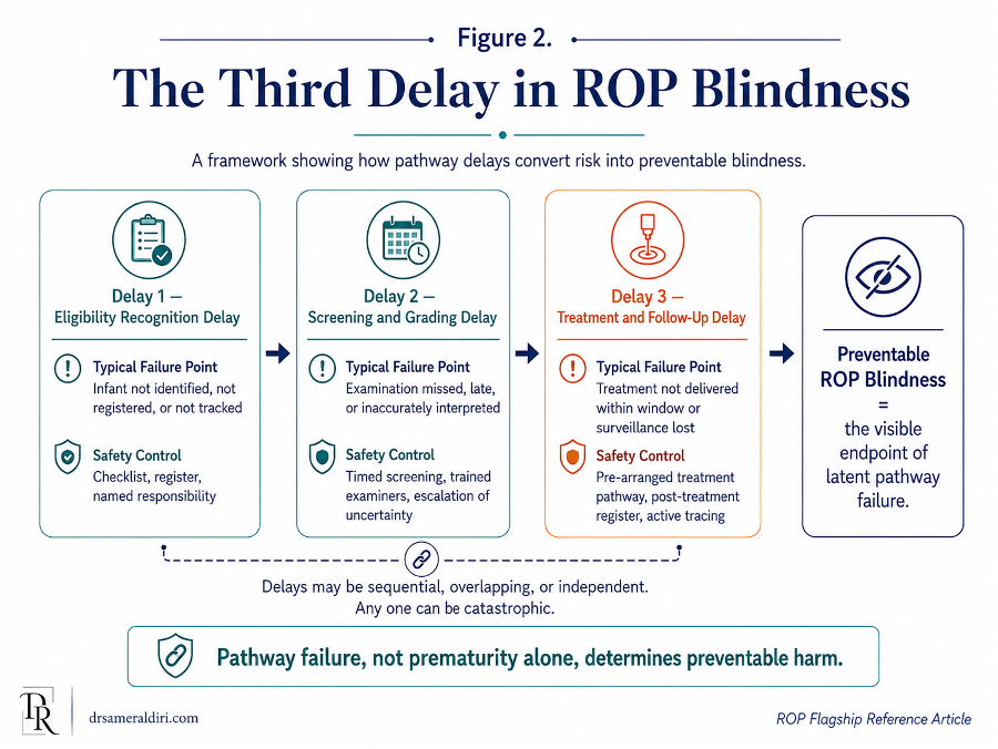
5.5 Data invisibility and underestimated burden
ROP programme failure can also be hidden by weak data. If treatment-requiring infants are not captured in a register, if treatment modality is not recorded, if retreatment is not measured, or if outcomes are not followed, leaders may believe the programme is safer than it is. The UK active-surveillance work is an important reminder that burden estimates can change when case ascertainment improves. What appears to be low incidence may sometimes reflect incomplete visibility rather than true low risk. [12]
5.6 Recurrence, retreatment, and the danger of premature reassurance
A pathway can also fail after apparently successful treatment. UK one-year outcome and retreatment data reinforce the need to avoid premature reassurance. Regression after laser or anti-VEGF therapy is encouraging, but it does not automatically close the pathway. Persistent activity, skip areas, late reactivation, recurrence, or longer-term visual consequences may still require specialist follow-up. [2, 13, 26, 28, 29]
This is particularly important where anti-VEGF therapy is used. Anti-VEGF may be clinically valuable in selected infants, especially with posterior or aggressive disease, but it increases the governance importance of prolonged surveillance. A system should not choose a treatment pathway unless it can also deliver the follow-up burden that pathway requires.
For this reason, every ROP programme should treat data quality as a safety function. The register should not exist only to count examinations. It should be able to show eligibility capture, screening timing, disease severity, treatment-requiring disease, treatment location, treatment modality, retreatment, post-treatment follow-up, visual and structural outcome where available, discharge from surveillance, and follow-up loss.
5.7 Programme response: treat delay as a safety event
A missed or delayed ROP step should not be treated as ordinary administration. It should trigger an active safety response. An overdue examination should be escalated. A poor-quality image should be repeated or referred. An unclear grading decision should receive senior review. A treatment-requiring case should activate the treatment pathway immediately. A missed post-treatment appointment should trigger active tracing, not passive waiting. [2, 7, 8, 9, 10, 11]
The aim is not to create fear. The aim is to create reliability. ROP prevention works when predictable failure points are converted into visible controls: registers, due lists, handover fields, escalation contacts, treatment-location plans, missed-appointment tracing, parent communication, and outcome audit. In ROP, the ethical task is to make the pathway strong enough that the baby is protected even when the system is busy, fragmented, or under pressure.
Part V has examined why ROP programmes fail and why preventable blindness often emerges from delay chains rather than from one isolated event. The next section turns this analysis into system design: how to build a safe ROP screening programme with registers, escalation routes, audit indicators, workforce planning, parent communication, and accountable leadership.
Part VI — Building a Safe ROP Screening System
Key Concept:
A safe ROP programme is defined not by the existence of screening alone, but by whether every eligible infant is reliably identified, examined, and followed, and whether infants with concerning or treatment-requiring disease are promptly escalated and treated across the full NICU-to-retina pathway. [1, 2, 3, 4]
Part V examined how Retinopathy of Prematurity (ROP) programmes fail. Part VI turns that analysis into system design. If preventable ROP blindness is often the endpoint of pathway failure, then prevention requires a pathway built deliberately enough to withstand pressure, transfer, staff rotation, parental stress, equipment gaps, and clinical urgency.
A safe ROP screening system is not created by a guideline alone. It is created when the guideline is translated into daily practice: when eligibility is checked, a register is active, examinations are scheduled, findings are documented, uncertain grading is escalated, treatment access is available, discharge is safe, parents are informed, and follow-up continues until risk is closed.
This is the practical meaning of ROP governance. Governance is not only policy. It is the set of responsibilities, controls, measurements, and escalation routes that make safe care more reliable than unsafe care.
6.1 The NICU-to-Retina Safety Chain
This article introduces the NICU-to-Retina Safety Chain as a systems-level construct describing the full pathway through which premature infants move from neonatal survival to vision protection. It begins before the first eye examination and continues beyond treatment or regression.[1, 3, 5, 6, 7, 14]
The chain includes:
- Neonatal survival and stabilisation
- Oxygen governance and systemic neonatal safety
- Eligibility recognition
- Entry into an ROP register
- Timed first screening examination
- Accurate classification and documentation
- Safe follow-up scheduling
- Urgent escalation of treatment-requiring disease
- Timely treatment where indicated
- Post-treatment surveillance
- Discharge and transfer safety
- Long-term paediatric ophthalmology follow-up where needed
This chain matters because no single link is sufficient alone. Excellent neonatal survival without screening leaves the infant exposed. Screening without accurate grading may miss treatment-requiring disease. Diagnosis without treatment access does not protect vision. Treatment without surveillance may allow reactivation or recurrence to be missed. Discharge without a secured appointment may interrupt the pathway at precisely the wrong moment.
A safe system therefore asks not only whether each link exists, but whether each link reliably connects to the next. The central governance question is: does the infant move through the pathway by design, or only because individual people remember to make it happen?
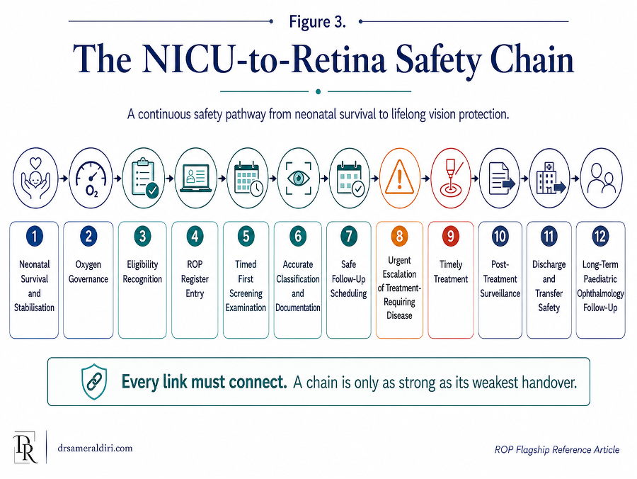
6.2 The ROP Blindness Prevention Cascade
To convert the safety chain into measurable programme steps, this article proposes the ROP Blindness Prevention Cascade. The cascade asks whether every infant who should enter the pathway actually reaches each protective stage. [6, 7, 30, 31, 32, 33]
The cascade can be summarised as:
Eligible → identified → registered → scheduled → screened on time → graded accurately → managed appropriately through repeat screening or urgent treatment escalation → treated within the required window if indicated → post-treatment or surveillance follow-up completed → safely discharged from ROP surveillance or formally transferred to longer-term care
This cascade is useful because it reveals where programme performance is lost. A hospital may report that many babies were screened, but the cascade asks deeper questions. Were all eligible babies identified? Were first examinations on time? Were findings classified accurately? Were follow-up intervals completed? Were treatment-requiring cases treated urgently? Were post-treatment visits completed? Were infants lost after discharge?
The purpose of the cascade is not to produce a perfect-looking chart. It is to make hidden failure visible. A drop-off at any point should trigger review. If infants are eligible but not registered, the eligibility system is weak. If infants are registered but not screened on time, scheduling or workforce capacity may be inadequate. If infants are screened but treatment is delayed, the escalation pathway may be unsafe. If infants are treated but not followed, post-treatment governance is incomplete.
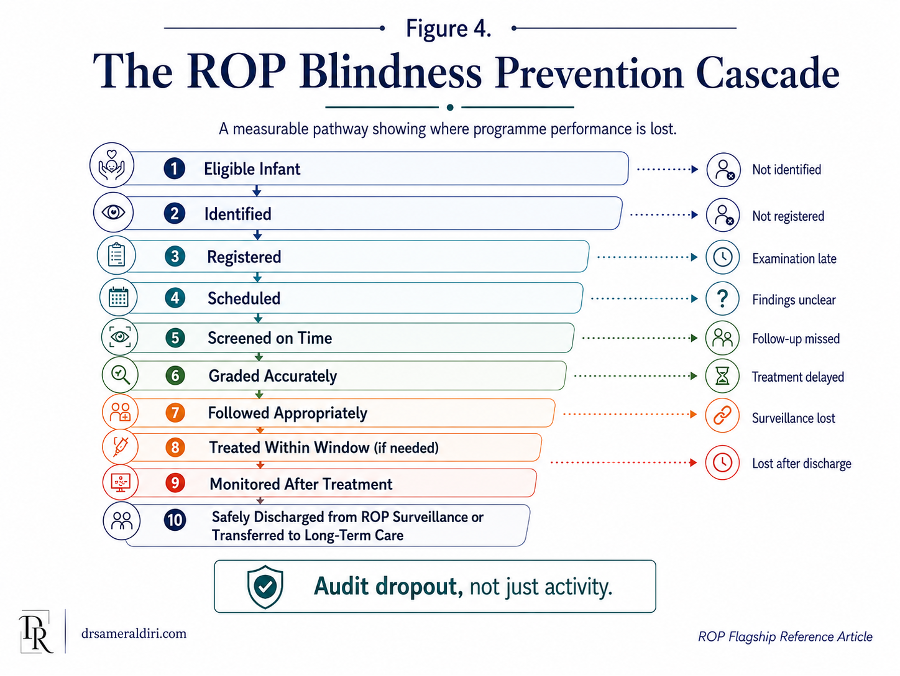
6.3 Governance roles: who owns each part of the pathway?
ROP prevention is multidisciplinary, but multidisciplinary care becomes unsafe when responsibility is shared so widely that no one is clearly accountable. A safe programme must define who owns each part of the pathway. [1, 30, 31, 34]
Neonatal teams usually hold the earliest responsibility. They identify at-risk infants, record gestational age and birth weight, recognise additional clinical risk, maintain neonatal stability, support oxygen governance, inform parents, and ensure that ROP status is considered before transfer or discharge.
Ophthalmology teams are responsible for retinal examination, image interpretation where applicable, disease classification, treatment decisions, follow-up intervals, escalation of severe disease, post-treatment surveillance, and long-term eye-care recommendations.
Nursing teams often provide the operational continuity that makes the pathway work. They may maintain registers, coordinate examinations, prepare infants, communicate with parents, support documentation, arrange appointments, and notice when a baby is approaching discharge or transfer before screening is complete.
Hospital leadership is responsible for ensuring that the pathway has the workforce, equipment, space, data systems, referral agreements, and quality oversight required to function. A hospital cannot delegate ROP safety entirely to individual clinicians if the system itself lacks the resources or structure needed to deliver care.
Regional or national health authorities have a wider responsibility. They define standards, support workforce planning, develop referral networks, monitor programme performance, reduce geographic inequity, and ensure that neonatal expansion is matched by screening and treatment capacity.
Parents are essential partners, but they should not be treated as the primary safety net. They need clear information, confirmed appointments, written instructions, and contact routes. Their role is to participate in care, not to compensate for an unreliable system.
6.4 The ROP register as the operational backbone
The ROP register is the operational backbone of a safe programme. Without a live register, the pathway depends too heavily on memory, informal referral, and fragmented documentation. [1, 3, 5, 14]
A useful ROP register should identify every infant who meets screening criteria or requires consideration for screening. It should record the infant’s gestational age, birth weight, postmenstrual age, neonatal unit, eligibility status, first examination due date, examination findings, follow-up interval, treatment status, transfer status, discharge status, and final outcome.
The register should not be a passive list. It should actively generate action. It should show which infants are due, overdue, awaiting treatment, post-treatment, transferred, discharged with pending follow-up, or ready for discharge from screening. It should allow the team to see risk before harm occurs.
A register may be electronic, paper-based, or hybrid. The format is less important than reliability. In a high-resource setting, an electronic register may link to alerts and appointment systems. In a resource-constrained setting, a carefully maintained paper register may still save sight if it is reviewed consistently, assigned to a responsible person, and integrated into ward rounds and discharge checks.
The most important principle is that no eligible infant should be invisible to the pathway.
6.5 Scheduling and due-list review
Scheduling is one of the most important controls in ROP safety. The first examination date should be calculated according to the relevant guideline and recorded in the register. Repeat examinations should be scheduled before the previous examination is considered complete. Treatment reviews should be booked before the infant leaves the treatment episode. [1, 2, 3, 4]
A safe programme should have a regular due-list review. This may be daily in large neonatal units or weekly in smaller services, depending on caseload and risk. The review should identify infants due for first examination, infants due for repeat examination, infants overdue, infants awaiting treatment, infants approaching discharge, and infants being transferred.
The due-list review should include both neonatal and ophthalmology teams, or at least ensure reliable communication between them. The purpose is to prevent time-critical care from being discovered late. An overdue ROP examination should not be found by accident. It should be visible on the system.
The due list is therefore more than a scheduling tool. It is a safety surveillance mechanism.
6.6 Escalation pathways for urgent disease
A screening system is incomplete unless it has an escalation pathway for treatment-requiring disease. The moment severe ROP is suspected or confirmed, the programme must be able to move from examination to action without unnecessary delay. [2, 7, 8, 9, 10, 11]
An escalation pathway should define:
- who must be contacted when treatment-requiring disease is found;
- how quickly senior ophthalmology review should occur;
- where treatment can be delivered;
- what equipment and neonatal support are required;
- how parents will be counselled and consent obtained;
- how urgent transfer will be arranged if treatment is not available locally;
- who confirms that treatment has occurred;
- how post-treatment follow-up is scheduled and tracked.
This pathway should be written before the emergency occurs. It should not be invented while a baby with severe ROP is waiting. Delays after diagnosis are particularly serious because the disease has already been recognised. The remaining question is whether the system can respond.
Escalation should also apply to uncertainty. Poor image quality, poor retinal view, suspected plus disease, possible aggressive ROP, posterior disease, or disagreement between graders should trigger senior review or referral. In ROP, uncertainty should move the infant toward protection, not passive delay.
6.7 Discharge and transfer controls
Discharge and transfer are high-risk transitions in the ROP pathway. A safe programme must therefore build ROP checks into both processes. [1, 3, 4]
Before discharge, the team should confirm:
- whether the infant is eligible for ROP screening;
- whether the first examination has been completed;
- what the last retinal findings were;
- whether follow-up is still required;
- when and where the next appointment will occur;
- who is responsible for the appointment;
- whether parents have received verbal and written information;
- whether contact details are correct;
- whether the discharge summary clearly states the ROP status and plan.
If follow-up is required after discharge, the appointment should be secured, not merely recommended. “Outpatient follow-up” is not enough unless the date, service, contact route, and urgency are clear.
Transfer requires the same discipline. The sending team should communicate ROP eligibility, screening status, last examination, next due date, treatment status, and urgency. The receiving team should confirm that it has accepted responsibility for continuing the pathway. ROP status should travel with the infant as clearly as oxygen requirement, medication, feeding plan, infection status, or respiratory support.
A premature infant should not lose retinal safety because they moved from one unit to another.
6.8 Parent communication as part of system design
Parent communication is often treated as a soft part of care. In ROP, it is a safety control.
Parents need to understand why screening is needed, why timing matters, what has been found, what the next appointment is, what treatment may involve, and who to contact if anything is unclear. This communication should be calm, practical, and repeated at key moments: before the first examination, after significant findings, before discharge, after treatment, and when long-term follow-up is needed. [1, 3, 4]
Written information is essential. Families may be exhausted, frightened, overwhelmed, or managing multiple neonatal appointments. A verbal explanation given during a stressful admission may not be remembered accurately. Written information should include the reason for screening, the importance of follow-up, appointment details, warning that delay may be harmful, and contact information.
However, parent communication should never become parent-blaming. The purpose is to make the pathway visible and supportive, not to transfer system responsibility onto the family. Parents should feel that they are part of a reliable care pathway, not that their baby’s vision depends on them chasing a fragmented system.
6.9 Audit indicators and KPIs
A safe ROP programme should measure what matters. Counting the number of babies screened is useful, but it is not enough. A programme can be busy and still unsafe if eligible infants are missed, examinations are late, findings are unclear, treatment is delayed, or follow-up is lost. [1, 3, 5, 14, 30, 31]
ROP audit should therefore include both activity and reliability indicators.
A practical ROP safety dashboard may include:
Table 5. ROP programme safety dashboard: domains, indicators, and safety meaning
| Domain | Indicator | Why it matters |
| Eligibility capture | Percentage of eligible infants entered into the ROP register | Measures whether the pathway starts reliably. |
| First examination timing | Percentage of first examinations completed within guideline timing | Measures whether screening begins on time. |
| Repeat examination reliability | Percentage of follow-up examinations completed within the recommended interval | Measures continuity of surveillance. |
| Documentation completeness | Percentage of examination records with zone, stage, vascular activity, follow-up interval, and next step documented | Measures whether findings are actionable. |
| Grading quality | Percentage of uncertain, poor-view, or image-quality-limited cases escalated for senior review | Measures whether uncertainty is managed safely. |
| Treatment escalation | Time from recognition of potentially treatment-requiring ROP to specialist treatment decision | Measures speed of response after serious disease is recognised. |
| Treatment delivery | Time from treatment decision to treatment completion | Measures whether the treatment window is protected. |
| Discharge safety | Percentage of infants discharged with ROP status and follow-up plan documented | Measures safety at transition home. |
| Transfer safety | Percentage of transferred infants with ROP status communicated and accepted by receiving unit | Measures continuity across institutions. |
| Post-treatment surveillance | Percentage of treated infants completing required post-treatment follow-up | Measures whether treatment is followed through. |
| Follow-up loss | Number and percentage of infants lost before safe discharge from ROP surveillance | Measures the pathway’s ability to retain infants until risk is closed. |
| Parent communication | Percentage of families receiving documented verbal and written ROP information | Measures whether parents are supported as partners. |
The most important indicators should be reviewed regularly by the programme team. Serious delays, missed examinations, treatment delays, and follow-up loss should be treated as safety events. The purpose of measurement is not to create bureaucracy. It is to detect weakness before a child is harmed.
6.10 Multidisciplinary accountability
ROP prevention requires a team, but a team still needs accountability. Multidisciplinary accountability means that each group understands its role and that the whole pathway is reviewed as one system. [30, 31, 33, 34]
A mature programme should have a named ROP lead or leadership group. This may include neonatal, ophthalmology, nursing, quality-safety, imaging, and administrative representation. The group should review register performance, overdue infants, treatment cases, transfer events, follow-up loss, equipment problems, training needs, and audit outcomes.
Accountability should be practical. It should answer:
- Who checks eligibility?
- Who maintains the register?
- Who calculates the first examination date?
- Who performs or arranges screening?
- Who confirms the next appointment?
- Who escalates severe or uncertain disease?
- Who arranges treatment or transfer?
- Who confirms post-treatment surveillance?
- Who ensures discharge and transfer safety?
- Who reviews missed or delayed care?
If these questions cannot be answered clearly, the pathway is not yet safe enough.
Accountability also means that the programme learns from failure. When a baby is screened late, treated late, transferred without retinal status, or lost to follow-up, the review should examine the pathway rather than only the individual event. The goal is to make the same failure harder to repeat.
6.11 Minimum safe system requirements
Not every health system has the same resources. Some programmes have electronic records, wide-field imaging, specialist ROP teams, and established referral networks. Others may rely on paper registers, periodic visiting ophthalmologists, and regional referral arrangements. The standard should therefore be realistic, but it must not be vague. [1, 3, 5, 14, 35, 36]
At minimum, a safe ROP system requires:
- clear eligibility criteria;
- a reliable register of eligible infants;
- a process for calculating first examination timing;
- access to trained examination or imaging capacity;
- standardised documentation of findings and next steps;
- defined follow-up intervals;
- urgent escalation for treatment-requiring or uncertain severe disease;
- a treatment pathway or referral agreement;
- discharge and transfer checks;
- parent communication and written information;
- active tracking of missed or overdue follow-up;
- audit of delays, treatment access, and outcomes.
These are not luxury components. They are the basic architecture of a preventable-blindness pathway. A programme may begin simply, but it must begin with reliability.
6.12 From safe system to mature programme
A basic ROP programme identifies eligible infants and arranges screening. A developing programme maintains a register and tracks follow-up. A functional programme audits timing, documentation, escalation, discharge, transfer, and treatment access. An advanced programme reviews outcomes, trains its workforce, monitors quality, reduces regional inequity, and integrates neonatal and ophthalmology data. An AI-enabled programme may use imaging, tele-ROP, or decision-support tools, but only within a governed pathway that ensures human oversight, treatment access, and accountability. [1, 3, 5, 14, 30, 31]
Maturity is not defined by technology alone. A programme with AI but weak follow-up is not mature. A programme with wide-field imaging but no treatment route is not mature. A programme with a guideline but no register is not mature. Maturity is defined by reliability across the full pathway.
The safest ROP systems are those that combine clinical expertise with operational discipline. They do not assume that good intentions will protect every infant. They build systems in which the next safe step is visible, expected, documented, and followed through.
Implementation maturity also depends on ownership and rhythm, not only on the presence of tools. Registers, dashboards, imaging systems and escalation pathways become protective only when a named team reviews them regularly, staff are trained to use them, overdue infants are acted on, and leaders have authority to resolve equipment, staffing, transfer and treatment barriers. [30, 31, 33, 34, 35, 36]
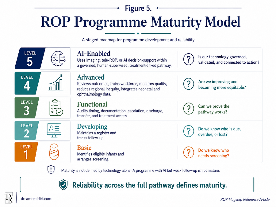
Table 6. ROP programme maturity model: basic, developing, functional, advanced, and AI-enabled
| Programme maturity level | Core characteristics | Main risks | Next development priority |
| Basic | Eligible infants are recognised and ROP screening is available, but the pathway may depend on individual referral, memory, or informal coordination. | Missed eligibility, late first examination, weak follow-up tracking, and inconsistent documentation. | Establish clear eligibility criteria, a live ROP register, first-examination timing, and basic follow-up tracking. |
| Developing | A register exists, screening is scheduled, and follow-up is tracked, but audit, escalation, discharge safety, and treatment access may still be inconsistent. | Overdue examinations, uncertain grading, discharge gaps, transfer communication failures, and delayed escalation. | Strengthen due-list review, standardised documentation, discharge and transfer checks, and urgent escalation rules. |
| Functional | The programme has eligibility capture, register review, screening, grading, documentation, treatment referral, discharge safety, and basic audit. | Variation in performance, workforce pressure, incomplete outcome review, and limited regional coordination. | Audit timing, treatment delay, follow-up loss, post-treatment surveillance, parent communication, and outcomes. |
| Advanced | The programme is multidisciplinary, audited, treatment-linked, regionally coordinated, and able to monitor quality, outcomes, workforce needs, and equity gaps. | Service expansion may outpace workforce, treatment capacity, or long-term follow-up systems. | Integrate neonatal and ophthalmology data, strengthen regional networks, expand training, and reduce inequity. |
| AI-enabled | Tele-ROP, wide-field imaging, or AI-supported tools are used within a governed clinical pathway with validation, human oversight, escalation, treatment access, data governance, and audit. | False reassurance, poor image quality, unvalidated tools, weak accountability, digital inequity, or detection without treatment access. | Ensure local validation, human accountability, equity monitoring, cybersecurity, and continuous review of AI-supported pathway performance. |
6.13 The leadership test
For hospital leaders and policymakers, the core question is not, “Do we have ROP screening?” The better question is, “Can we prove that every eligible infant moves safely through the full pathway?” [1, 3, 5, 14]
This requires evidence. A leader should be able to ask for the ROP register, the overdue list, the number of infants screened on time, the number treated, the treatment delay interval, the follow-up loss rate, the discharge safety process, the transfer handover process, the equipment status, the workforce plan, and the audit record.
If these cannot be shown, the programme may exist in name but not yet in dependable practice.
ROP prevention is therefore a leadership test. It tests whether a health system can connect neonatal care, ophthalmology, nursing, data, referral, treatment, parent communication, and long-term follow-up into one coherent pathway. It tests whether the system notices risk before harm occurs. It tests whether survival is matched by protection.
The final message of Part VI is direct: a safe ROP programme does not rely on heroic individuals remembering every step. It relies on a designed pathway that makes preventable blindness harder to occur.
6.14 From national surveillance to local operational dashboards
The UK surveillance literature also shows what local programmes should be able to know about themselves. At hospital or regional level, leaders should be able to identify how many infants were eligible, how many were screened on time, how many developed treatment-requiring ROP, where treatment occurred, what treatment modality was used, whether neonatal or anaesthetic support was required, how many infants required retreatment, and what structural or visual outcomes were documented. These data should not be gathered only for research. They should be used to manage service capacity, identify bottlenecks, and prevent delay. [12, 13, 15, 25]
A practical ROP dashboard should therefore combine screening-process metrics with treatment and outcome metrics. Counting examinations alone is insufficient. The dashboard should also show treatment delay intervals, post-treatment review completion, recurrence or reactivation tracking, discharge from ROP surveillance, and longer-term paediatric ophthalmology follow-up where indicated. This is how a register becomes a safety instrument rather than a static list.
Part VI has described how to build a safe ROP screening system. The next section turns to the governance of AI in ROP screening: how tele-ROP, wide-field imaging, and AI-supported detection may extend capacity, but only when they are clinically validated, ethically governed, linked to treatment access, and embedded within human accountability.
Part VII — The Governance of AI in ROP Screening
Key Concept:
AI may extend screening capacity, but it must remain governed by human accountability, validation, data quality, escalation pathways, and treatment access. [35, 38, 39, 40, 41, 42, 43, 44, 45, 46]
Artificial intelligence has entered the conversation about Retinopathy of Prematurity (ROP) because the problem is both clinically urgent and operationally difficult. Many health systems do not have enough specialists to examine every at-risk premature infant at the right time. Some neonatal units are geographically distant from paediatric retinal expertise. Some programmes struggle with workforce shortages, inconsistent grading, poor documentation, referral delay, and follow-up loss. In this context, tele-ROP, wide-field retinal imaging, and AI-supported analysis may help extend capacity and improve consistency.
However, AI does not remove the core responsibility of the health system. It cannot replace a screening register. It cannot counsel parents. It cannot create treatment access where none exists. It cannot guarantee that an infant with severe disease will be transferred, treated, and followed. It cannot carry professional accountability for a child’s sight.
The central question is therefore not whether AI is impressive. The central question is whether AI is safe, validated, governed, clinically integrated, equitable, and connected to action.
7.1 Why AI and tele-ROP matter
ROP screening is time-critical, specialist-dependent, and vulnerable to capacity gaps. A premature infant may be in a neonatal unit where an experienced ROP examiner is not available every day. Specialist ophthalmologists may need to travel between hospitals. Rural or remote neonatal units may have limited access to paediatric retinal expertise. In some settings, the number of premature infants requiring screening is increasing faster than the trained workforce. [1, 3, 5, 14, 35, 36]
Tele-ROP developed as one response to this challenge. In a tele-ROP model, trained personnel capture retinal images, usually using wide-field retinal imaging equipment, and the images are reviewed by qualified experts at another location. This can extend specialist interpretation beyond the physical presence of an ophthalmologist at every bedside.
AI may add another layer. It may support detection of disease activity, assessment of vascular severity, identification of plus disease or pre-plus features, image quality assessment, risk stratification, triage, workflow prioritisation, and decision support. In principle, this could help programmes identify high-risk infants earlier, reduce variation between graders, support overstretched services, and focus urgent specialist attention where it is most needed.
But the promise of AI should not obscure the nature of ROP care. ROP screening is not only image interpretation. It is a pathway from neonatal admission to retinal examination, diagnosis, treatment, discharge safety, and long-term follow-up. AI may assist one or more steps in that pathway, but it does not replace the pathway itself.
7.2 Tele-ROP as the foundation for AI-supported screening
AI in ROP is usually built on image-based screening. This means that the quality of AI-supported care depends first on the quality of the imaging pathway. [1, 3, 5, 14, 35, 36]
A safe tele-ROP system requires trained image acquisition, appropriate equipment, clear eligibility criteria, timely imaging, secure image transfer, qualified interpretation, structured reporting, escalation rules, and treatment access. If any of these elements are weak, adding AI may not improve safety. It may simply automate part of an unreliable system.
Tele-ROP can be powerful because it separates image capture from expert interpretation. A neonatal unit may capture images locally while an experienced ROP specialist reviews them remotely. This can reduce travel burden, extend access, and support regional networks. It can also create a digital record that can be reviewed, audited, taught from, and compared over time.
However, tele-ROP also introduces new risks. Images may be poor quality. Peripheral retina may not be captured adequately. The baby may be too unstable for imaging. The imaging session may be delayed. The reviewer may not receive images in time. Reports may not be acted upon. A recommendation for urgent treatment may still fail if no treatment pathway exists.
Tele-ROP should therefore be treated as a clinical service, not as a technology purchase. Buying an imaging device does not create a safe tele-ROP programme. Safety depends on people, process, quality assurance, escalation, and accountability.
7.3 Where AI can support the ROP pathway
AI may support ROP screening in several ways.
First, it may help assess vascular severity. One of the most challenging aspects of ROP classification is judging plus disease and pre-plus disease. These assessments can vary between clinicians, particularly in borderline cases. AI-supported vascular analysis may help provide more consistent severity scoring or highlight images that need urgent review. [38, 39, 40, 44]
Second, AI may assist with triage. In a busy programme, AI could help prioritise images that appear more concerning, allowing expert reviewers to focus quickly on infants at highest risk. This does not mean that low-risk infants can be ignored. It means that workflow may be organised more intelligently when the system is under pressure.
Third, AI may support image quality control. If images are blurred, incomplete, poorly centred, or insufficient to assess the retinal periphery, AI may help flag that the examination is inadequate. This is important because a poor-quality image should never be treated as a normal result.
Fourth, AI may support risk stratification. By combining retinal image features with clinical data such as gestational age, birth weight, postmenstrual age, oxygen exposure, disease trajectory, or prior findings, future systems may help predict which infants require closer follow-up. Such tools would need careful validation before clinical use.
Fifth, AI may support audit and programme improvement. Aggregated data may help identify variation in grading, delayed review, poor image quality, overdue follow-up, or referral bottlenecks. In this role, AI may support quality assurance rather than direct diagnosis.
Each of these uses has potential value. Each also carries risk if implemented without governance.
7.4 What AI must not be asked to do
AI should not be asked to compensate for an unsafe pathway. If eligible infants are not identified, AI cannot screen them. If images are not captured, AI cannot interpret them. If image quality is poor, AI output may be unreliable. If reports are not reviewed, AI cannot act on them. If treatment is unavailable, AI cannot prevent blindness. If follow-up is not tracked, AI cannot ensure that the child remains safe. [44, 45, 46, 57]
AI should not be used to create false reassurance. A low-risk AI output should not override clinical concern, poor image quality, incomplete retinal views, or worsening systemic context. A system that accepts AI reassurance without checking whether the image was adequate, the infant was eligible, the timing was correct, and follow-up was secured is not practising safe AI governance.
AI should not be deployed as a substitute for workforce planning. It may support workforce capacity, but it cannot replace trained clinicians, imaging staff, neonatal teams, treatment specialists, and programme coordinators. If leaders use AI to avoid building human capacity, the programme may become more fragile rather than safer.
AI should not be introduced without a clear answer to a simple question: who is accountable if the output is wrong, ignored, delayed, or not acted upon?
7.5 Validation before adoption
AI tools used in ROP screening must be validated before they are trusted in clinical pathways. Validation means more than a high accuracy number in a research paper. It means demonstrating that the system performs safely in the population, imaging conditions, disease spectrum, equipment environment, and workflow where it will actually be used. [41, 42, 43, 46]
An AI model trained on images from one region, one device, one imaging protocol, or one disease distribution may not perform equally well elsewhere. Premature infants differ across settings. Image quality varies. Disease prevalence varies. Examiner and imager experience varies. Treatment thresholds and referral pathways may vary. A tool that performs well in a curated research dataset may perform differently in a busy neonatal unit.
Validation should also focus on clinically meaningful outcomes. Retrospective development, test-set performance and external-validation studies can establish diagnostic performance under defined conditions, but they do not by themselves demonstrate prospective clinical utility, safe workflow integration, improved patient outcomes, or regulatory readiness. Before routine clinical reliance, an AI tool should therefore undergo proportionate prospective evaluation in its intended setting, assessment within the complete screening-to-treatment pathway, review against applicable medical-device and data-governance requirements, and continuing post-deployment monitoring for performance drift, bias, workflow failure and unintended harm. [41, 42, 43, 45, 46, 57]
External validation should be treated as a governance requirement, not an academic ornament. A model may perform well in development data and still behave differently across imaging devices, ethnic and geographic populations, disease prevalence, image-acquisition skill, retinal pigmentation, field of view, and neonatal case mix. Multi-country validation, low- and middle-income setting studies, and smartphone-based telescreening evidence are encouraging, but they reinforce the same principle: AI must be locally monitored, clinically accountable, and connected to timely human review and treatment access.
7.6 Data quality and imaging governance
AI quality depends on data quality. In ROP, data quality begins at the bedside.
A safe imaging pathway should define who is trained to capture images, how image quality is assessed, which retinal fields are required, how incomplete imaging is documented, how quickly images must be uploaded or reviewed, and what happens when images are inadequate. Poor image quality should trigger repeat imaging, bedside examination, senior review, or referral according to risk. It should not be treated as a negative screen. [35, 36, 44, 45, 46]
Image datasets used for AI development and monitoring should be labelled carefully. Labels should be based on appropriate clinical expertise, standardised terminology, and clear definitions. If labels are inconsistent, the AI may learn inconsistency. If plus disease is variably graded by humans, the model may reproduce or amplify that variability unless the training process is carefully governed.
Data governance should also address privacy, consent, storage, transfer, access, audit trails, cybersecurity, and data retention. Retinal images of premature infants are sensitive health data. They should be handled with the same seriousness as any other neonatal medical record.
AI governance therefore begins before the algorithm runs. It begins with image acquisition, data stewardship, clinical labelling, secure infrastructure, and quality assurance.
7.7 Human oversight and clinical accountability
Human oversight is essential in AI-supported ROP screening. The infant is not being cared for by an algorithm. The infant is being cared for by a clinical system that may use algorithmic support. [44, 45, 46]
A safe model of oversight should define who reviews AI output, who can override it, who confirms severe disease, who communicates with parents, who arranges treatment, and who is responsible for follow-up. AI output should be integrated into a clinical workflow where action is assigned, documented, and traceable.
Human oversight should not be passive. It should not mean that a clinician simply glances at an AI label after the system has effectively made the decision. Oversight should be meaningful: clinicians must understand the tool’s intended use, limitations, failure modes, uncertainty signals, image-quality requirements, and escalation rules.
Clinical accountability also means that the programme must define what happens when AI and clinician judgement disagree. If the AI output is reassuring but the clinician is concerned, the pathway should favour safety. If the AI output is concerning but the clinician is uncertain, the pathway should trigger review rather than dismissal. If image quality is inadequate, the result should not be treated as reliable.
In ROP, human accountability is not a symbolic safeguard. It is the mechanism by which AI output is converted into safe clinical action.
7.8 Bias, equity, and local performance
AI can reduce inequity if it extends access to screening expertise in underserved settings. It can also worsen inequity if it performs less well in the very populations that most need support. [41, 43, 44, 45, 46, 57]
Bias may enter an AI system through training data, image devices, image quality, disease prevalence, population characteristics, or clinical labels. A tool trained mostly on high-quality images from well-resourced settings may be less reliable in clinics using lower-cost devices, less experienced imagers, or different disease patterns. A system trained in one population may not perform equally well in another.
Equity therefore requires more than making AI available. It requires proving that AI works safely across the populations and settings where it is deployed. It also requires ensuring that positive or uncertain results lead to real care. An AI system that detects risk in a remote unit but cannot connect the baby to treatment may expose inequity rather than solve it.
AI should therefore be monitored for differential performance. Programmes should ask whether sensitivity, specificity, image-quality failure rates, false negatives, false positives, escalation delays, treatment completion, and follow-up loss differ by hospital, geography, device, gestational age, birth weight, ethnicity where ethically and legally recorded, socioeconomic context, or transfer status.
The equity standard is not that every site has the same technology. The standard is that every eligible infant has a safe pathway from risk detection to treatment and follow-up.
7.9 AI must be linked to treatment access
The greatest danger in AI-supported ROP screening is the illusion that detection alone is prevention. It is not.
An AI system may identify a baby at high risk. It may highlight vascular abnormality. It may support recognition of plus disease. It may recommend urgent review. But unless the programme has a functioning escalation and treatment pathway, the baby remains at risk. [35, 37, 44, 45, 46, 57]
Before deploying AI, leaders should ask whether the system can act on what AI finds. Is there a treating ophthalmologist or referral centre? Is laser available? Is anti-VEGF therapy governed by protocol? Is neonatal support available for treatment? Can urgent transfer be arranged? Can parents be counselled quickly? Can post-treatment surveillance be tracked? Can late reactivation after anti-VEGF therapy be detected?
If the answer is no, AI may increase the visibility of risk without reducing the risk itself. In some cases, this may still be useful because it exposes a gap. But it should not be presented as a complete prevention solution.
AI in ROP is safe only when detection is connected to decision, decision to treatment, treatment to surveillance, and surveillance to long-term vision care.
7.10 Legal, ethical, and professional responsibility
AI-supported ROP screening raises legal and ethical questions that must be addressed before implementation. Who is responsible for the output? Who is responsible for acting on it? What happens if the algorithm misses severe disease? What happens if image quality is inadequate but the system produces a reassuring result? What happens if an alert is generated but not reviewed? What happens if treatment access is unavailable? [44, 45, 46, 57]
These questions should not be left to individual clinicians at the bedside. They should be addressed through institutional policy, professional standards, consent processes, risk management, data governance, and audit. The programme should define the intended use of the AI tool, the limits of its use, the required human oversight, the escalation process, and the documentation requirements.
Governance should also be aligned with the jurisdiction in which the tool is used, including medical-device regulation where applicable, data-protection requirements, image-storage and sharing rules, procurement standards, professional indemnity arrangements, and local policies on consent, accountability and incident reporting. A tool that is technically promising is not automatically deployment-ready unless these regulatory and medico-legal conditions are explicit. [45, 46, 57]
Parents should be informed when AI-supported tools are used in their baby’s screening pathway, in language that is understandable and reassuring. The explanation should make clear that AI is a support tool and that clinical responsibility remains with the healthcare team. Consent processes should be aligned with local law, institutional policy, and the nature of the technology being used.
Ethically, AI should strengthen care rather than dilute responsibility. It should make the pathway safer, clearer, and more reliable. It should not create ambiguity about who is responsible for the child.
7.11 Implementation in resource-constrained settings
AI and tele-ROP are often discussed as solutions for resource-constrained settings. This is understandable. Where specialists are scarce and neonatal units are geographically dispersed, image-based screening and remote review may extend access. AI may further support triage and consistency. [1, 3, 5, 14, 18, 19]
However, resource-constrained settings also face the highest implementation risk. Imaging devices may be expensive or difficult to maintain. Internet connectivity may be unreliable. Staff turnover may interrupt training. Referral pathways may be weak. Treatment centres may be distant. Families may face transport and cost barriers. Data governance systems may be underdeveloped.
For this reason, AI implementation should be staged. A programme may first need to establish eligibility criteria, a register, imaging workflow, trained personnel, referral agreements, and treatment access. AI can then be added to strengthen the pathway, not to replace the foundations.
A simple but reliable paper register may be safer than an AI tool embedded in a chaotic pathway. A well-governed tele-ROP network with human expert review may be safer than an autonomous AI system without treatment access. Technology should be introduced at the maturity level the system can safely support.
The goal is not to deny innovation to lower-resource settings. The goal is to make innovation safe, useful, and equitable.
Table 7. AI and tele-ROP governance: opportunities, risks, safeguards, and accountability
| AI or tele-ROP component | Opportunity | Key risk | Required safeguard | Governance question |
| Wide-field retinal imaging | Extends screening access and creates a reviewable digital record. | Poor-quality or incomplete images may miss peripheral or posterior disease. | Imager training, image-quality standards, repeat imaging or bedside examination if inadequate. | Who confirms image quality and acts when images are inadequate? |
| Tele-ROP expert review | Allows remote specialist interpretation. | Review may be delayed or reports may not be acted upon. | Turnaround-time standards, structured reports, escalation alerts, audit. | Who ensures that reports lead to action? |
| AI vascular severity assessment | May support more consistent assessment of plus or pre-plus disease. | Model may perform differently across populations, devices, or image quality. | External validation, local testing, continuous monitoring, human review. | Has the model been validated in the population and setting where it is used? |
| AI triage | May prioritise high-risk infants for urgent review. | Low-risk output may create false reassurance. | Clear intended use, safety thresholds, override rules, audit of false negatives. | What happens when AI output and clinical concern disagree? |
| AI image-quality assessment | May flag unreadable or incomplete images. | Inadequate images may still be treated as reassuring if workflow is weak. | Mandatory repeat imaging or clinical examination when quality is insufficient. | Are inadequate images escalated rather than closed as negative? |
| AI risk prediction | May support follow-up planning and resource prioritisation. | Risk models may not generalise across populations or clinical contexts. | Prospective validation, subgroup analysis, clinician oversight. | Does risk prediction improve safety without increasing inequity? |
| Automated reporting | May standardise documentation and reduce omissions. | Over-reliance on templated output may hide uncertainty. | Clinician confirmation, mandatory uncertainty fields, audit trail. | Does the report clearly state next step, urgency, and responsibility? |
| Remote network governance | May link neonatal units to regional expertise. | Detection without treatment access may expose risk without solving it. | Referral agreements, treatment pathways, transfer protocols, post-treatment tracking. | Can every positive or urgent case reach treatment within the required window? |
| Data storage and transfer | Enables longitudinal comparison, audit, and training. | Privacy breach, unclear consent, weak cybersecurity, inappropriate reuse. | Data governance policy, secure systems, access controls, consent framework. | Who owns, accesses, protects, and audits the data? |
| AI-enabled programme monitoring | May identify delays, quality gaps, and missed follow-up. | Dashboard data may be incomplete or ignored. | Regular governance review, named responsibility, action tracking. | Who reviews the data and closes identified gaps? |
7.12 Minimum governance requirements before AI deployment
Before AI is introduced into an ROP screening programme, the following minimum requirements should be in place:
- clear eligibility criteria and a functioning ROP register;
- trained imaging or examination personnel;
- defined image-quality standards;
- secure image storage and transfer;
- validated AI performance for the intended use;
- local performance review before full clinical reliance;
- human oversight and override rules;
- clear documentation of AI output and clinical decision-making;
- escalation pathways for positive, uncertain, or poor-quality results;
- access to treatment or a formal referral route;
- post-treatment and long-term follow-up tracking;
- parent communication and consent processes;
- privacy, cybersecurity, and data governance policies;
- audit of false negatives, false positives, delays, missed follow-up, and outcomes;
- named clinical and institutional accountability.
These requirements are not barriers to innovation. They are what make innovation clinically trustworthy.
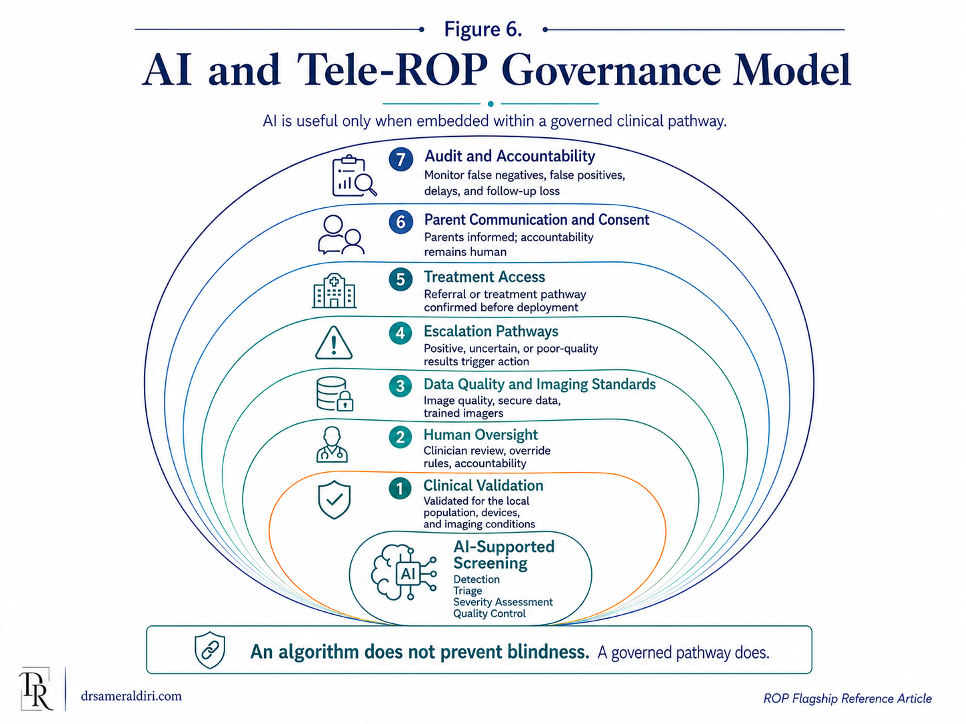
7.13 From technology adoption to governed innovation
The most important risk in AI-supported ROP screening is not that technology will fail completely. It is that technology will work partially, appear impressive, and still leave infants unsafe because the pathway around it is weak. [44, 45, 46, 57]
A programme may have AI but no reliable register. Imaging but no rapid interpretation. Triage but no treatment access. Automated output but no parent communication. Alerts but no escalation. Data but no governance. Innovation becomes unsafe when it gives the appearance of control without the substance of accountability.
Governed innovation is different. It asks what problem the technology is solving, how the tool has been validated, where it fits in the pathway, who is responsible for acting on its output, how uncertainty is managed, how parents are informed, how treatment access is secured, and how performance is monitored over time.
AI should therefore be introduced as a strengthening tool within a mature or maturing ROP pathway. It should support clinicians, not displace responsibility. It should extend access, not create a two-tier system. It should improve consistency, not hide uncertainty. It should accelerate escalation, not generate reports that no one acts upon.
The final message of Part VII is clear: AI can help ROP screening only when it is clinically validated, ethically governed, operationally integrated, and linked to treatment and follow-up. In ROP, an algorithm does not prevent blindness. A governed pathway using the right tools at the right time prevents blindness.
Part VII has examined the governance of AI in ROP screening. The next section turns to the comparative policy landscape: how different health systems organise screening, treatment, accountability, and implementation, and what global programmes can learn from established and emerging models.
Part VIII — Comparative Policy Landscape
Key Concept :
The strongest ROP policies do more than define screening criteria; they assign responsibility, protect follow-up, secure treatment access, and make programme performance auditable. [1, 3, 5, 14, 30, 31]
Retinopathy of Prematurity (ROP) policy varies across countries, but the central policy question is the same everywhere: how does a health system ensure that every eligible premature infant is identified, screened, followed, treated when needed, and protected from avoidable blindness?
A comparative policy landscape is useful not because one country’s model can simply be copied into another. Health systems differ in neonatal survival patterns, workforce capacity, geography, financing, digital infrastructure, referral networks, and treatment availability. A guideline that works in one setting may fail in another if the pathway around it is weak.
The purpose of comparison is therefore not to rank systems. It is to identify design principles.
The comparative evidence base should therefore be read selectively and practically. The UK contributes detailed guidance and national surveillance evidence. The United States contributes professional society guidance and large-system screening data. Australasia illustrates policy in a mature neonatal network environment. India demonstrates the operational value of wide-field imaging, tele-screening, non-physician acquisition and structured national guidance at scale. South American and South African literature shows how regions facing survival gains and resource variation adapt prevention, screening and treatment programmes to local realities. The common lesson is not one universal threshold. It is locally accountable implementation. [24]
Mature ROP policy must answer five questions:
Who is eligible for screening?
Who is responsible for ensuring that screening happens?
How is timing protected?
How is treatment access secured?
How is performance audited?
A policy that answers only the first question is incomplete. Screening criteria matter, but they do not prevent blindness by themselves. The real test is whether policy becomes an accountable pathway.
8.1 From guideline to governance
ROP policy usually begins with screening criteria. These criteria define which infants should be examined, using gestational age, birth weight, postmenstrual age, and sometimes additional clinical risk factors. This is essential because the population at risk must be clearly identified. [1, 3, 5, 14]
However, screening criteria are only the entry point. A safe ROP policy must also define timing of the first examination, repeat examination intervals, documentation standards, discharge and transfer responsibilities, treatment escalation, post-treatment surveillance, parent communication, and audit.
This distinction is important. A country may have an excellent written guideline, but infants may still be missed if the guideline is not embedded into neonatal practice. A hospital may know the criteria, but fail to maintain a live register. A screening programme may perform examinations, but fail to secure treatment access. A neonatal network may transfer infants, but fail to transfer retinal status. A parent may be told to attend follow-up, but receive no appointment.
ROP policy therefore has two levels. The first is the guideline: what should happen. The second is governance: how the system ensures that it actually happens.
8.2 United Kingdom: national guidance with separated screening and treatment responsibilities
The United Kingdom provides an important example of structured national ROP guidance. The UK approach separates screening and treatment into companion guidance, with screening guidance led through paediatric and neonatal collaboration and treatment guidance led through ophthalmology. This separation is useful because screening and treatment require different operational responsibilities, while still belonging to one continuous safety pathway. [1, 3, 5, 14, 12, 13]
The UK model is notable for several reasons. First, it treats ROP as a largely preventable cause of childhood visual disability and places emphasis on screening eligible infants at the right time. Second, it provides practical resources that support implementation, including screening documentation tools, parent and carer information, and calculation support for timing screening appointments. Third, it recognises that screening and treatment guidance must be updated as evidence evolves.
The policy lesson from the UK is not only the specific eligibility threshold. The deeper lesson is that national guidance must be translated into local neonatal and ophthalmology systems. Neonatal units need processes to identify eligible infants. Ophthalmology services need capacity to examine, classify, and escalate disease. Hospitals need discharge and transfer controls. Networks need referral arrangements for treatment. Parents need clear written information.
The UK model also illustrates a wider point: policy should not be static. Screening criteria and treatment approaches require periodic review, especially when new evidence suggests that some infants may fall outside previous thresholds or when treatment practice changes. A safe policy system must therefore have a mechanism for review, revision, and implementation support.
The UK experience is especially valuable because it links guidance with surveillance evidence. UK studies involving Adams, Bunce, Xing, Dahlmann-Noor and colleagues have examined treatment trends, screening burden, retreatment rates, one-year structural and visual outcomes, and neonatal anaesthetic or treatment-location arrangements. Together, these studies show that a mature ROP policy landscape must govern the whole pathway: who is screened, who is treated, how treatment is delivered, how retreatment is detected, and how outcomes are followed. [12, 13, 14, 15]
8.3 United States: professional society policy and programme responsibility
The United States model is shaped by professional society policy guidance, including paediatrics, ophthalmology, paediatric ophthalmology, and orthoptic expertise. This model emphasises the need to identify at-risk premature infants, perform examinations at proper times and intervals, and detect treatment-requiring ROP before destructive changes occur.[3]
The strength of this approach is its explicit recognition that severe ROP must be diagnosed in a timely fashion to be treated effectively. It treats ROP screening as a programme with defined attributes, not merely an isolated examination. It also highlights the sequential nature of ROP: infants may require repeated examinations because the disease can develop, progress, regress, or require treatment over time. [1, 3, 5, 14]
The US approach is also important because it places responsibility on the clinical system to ensure that screening and follow-up are arranged. In practice, this means that neonatal teams, ophthalmologists, hospital systems, and discharge planners must work together. The infant’s pathway must not become unsafe at discharge, transfer, or outpatient transition.
The policy lesson from the United States is that professional standards can define clinical expectations, but institutional implementation determines whether those expectations protect every infant. A policy statement can describe who should be screened and when. A hospital must still build the register, scheduling system, documentation process, parent communication pathway, treatment access, and follow-up tracking required to make the policy real. [3]
8.4 Australasia: guideline-based care with emphasis on local protocol and team responsibility
The Australasian model, represented by RANZCO guidance for Australia and New Zealand, provides a useful example of combining national professional guidance with local protocol responsibility. It recognises ROP as a potentially blinding disease in premature infants and frames ROP rates and outcomes as a possible proxy measure of neonatal-care standards.[1, 3, 4, 23, 24, 37]
This framing is important. It places ROP not only within ophthalmology, but also within neonatal quality. If ROP outcomes reflect neonatal care, oxygen governance, screening reliability, treatment access, and follow-up, then ROP becomes a marker of system performance rather than only a specialist retinal diagnosis.
The Australasian guidance also recognises team-based management. ROP care requires ophthalmologists, neonatologists, nurses, other staff, and parents to work together. It also supports the development of local screening protocols within neonatal units. This matters because national guidance must be adapted into a workable local pathway that reflects local staffing, geography, equipment, referral networks, and neonatal caseload.
The policy lesson from Australasia is that professional guidance should set standards while allowing local protocols to define how those standards are operationalised. The risk, however, is that local variation must not become local unreliability. Every local pathway still needs eligibility capture, documentation, follow-up timing, treatment escalation, discharge safety, transfer communication, and audit.
8.5 India: broader eligibility, public-health scale, and implementation innovation
India provides a particularly important policy and implementation case because it reflects the reality of a large middle-income country with major neonatal survival gains, diverse care settings, urban-rural variation, workforce constraints, and significant child-eye-health needs. [18, 19, 20, 21, 22, 23]
Indian ROP guidance has often used broader screening eligibility than many high-income settings because larger and more mature infants may still be at risk in some neonatal environments. This reflects a key principle: screening criteria should be appropriate to the local epidemiology, neonatal-care quality, oxygen governance, and survival patterns. A threshold that is safe in one system may be too narrow in another.
India is also important because it has generated implementation models that address workforce and geographic barriers. The Karnataka Internet Assisted Diagnosis of Retinopathy of Prematurity programme, commonly known as KIDROP, is an example of a tele-ROP model designed to reach underserved rural and semi-urban neonatal units. The model used wide-field imaging, trained non-physician imagers or graders, remote expert interpretation, and structured reporting to extend screening capacity beyond specialist hospital centres.
The policy lesson from India is that ROP prevention cannot rely only on tertiary centres. In a large health system, preventable blindness may occur far from specialist expertise. Public-health models must therefore use task-sharing, telemedicine, regional networks, transport planning, parent communication, referral agreements, and treatment access to bring the pathway closer to the infant.
India also shows that ROP policy must be implementation-focused. Written eligibility criteria are necessary, but they must be linked to neonatal-unit mapping, workforce training, oxygen governance, imaging capacity, treatment networks, data collection, and accountability. In a system of this scale, ROP prevention must be treated as a programme, not only a guideline.
8.6 Resource-constrained and middle-income settings: adapting principles, not copying thresholds
Resource-constrained and middle-income settings face a distinctive policy challenge. Neonatal survival may improve faster than the systems required to protect vision. This creates the Survival–Vision Paradox at policy level: more premature infants survive, but screening and treatment infrastructure may not yet be strong enough to protect every eligible infant. [1, 3, 5, 14, 18, 19]
In these settings, direct transplantation of high-income screening thresholds may be unsafe if local neonatal-care conditions differ. Broader eligibility may be needed where larger or more mature infants remain at risk because of variable oxygen monitoring, sepsis, poor weight gain, unstable neonatal courses, or inconsistent neonatal quality. Conversely, excessively broad criteria without sufficient workforce may overwhelm services and reduce reliability. Policy must therefore balance sensitivity, feasibility, local epidemiology, and programme capacity.
The most important policy requirement is not simply choosing a threshold. It is building a pathway that can deliver on the threshold chosen. If a system decides that a wide group of infants should be screened, it must provide the workforce, equipment, scheduling, follow-up, treatment access, and data systems required. If it cannot yet do so, phased implementation may be needed, with high-risk infants protected first while capacity is expanded.
Resource-constrained policy should therefore prioritise minimum safe-system requirements: clear eligibility criteria, a live register, defined screening timing, trained examiners or imagers, referral pathways, treatment access, discharge and transfer controls, parent communication, and audit of missed or delayed care. Technology may help, but it cannot substitute for these foundations.
8.7 Screening mandates and accountability
One of the most important differences between ROP policy environments is whether screening is treated as mandatory, expected, recommended, or optional. The language matters because it shapes accountability. [1, 3, 5, 14, 30, 31]
If screening is treated as optional, responsibility becomes fragile. Neonatal teams may request it inconsistently. Eye services may respond when asked but not actively track eligible infants. Parents may be told to follow up but not supported by a reliable system. Missed screening may be viewed as unfortunate rather than as a safety failure.
If screening is treated as a formal neonatal safety requirement, the system behaves differently. Eligibility is checked. Registers are maintained. First examinations are scheduled. Overdue infants are visible. Discharge cannot close the pathway without a plan. Transfer requires retinal status. Treatment-requiring disease triggers escalation. Follow-up loss is investigated.
The policy lesson is clear: ROP screening should not be left to informal referral culture. It should be embedded into neonatal governance. The infant should enter the pathway because they meet criteria, not because someone happens to remember to ask.
8.8 Audit: the difference between policy presence and policy performance
A policy exists on paper. A programme exists in practice. Audit is the bridge between the two.
Good ROP audit should not only count how many infants were screened. It should measure whether all eligible infants were captured, whether first examinations occurred within the required window, whether repeat examinations occurred at safe intervals, whether documentation was complete, whether uncertainty was escalated, whether treatment-requiring disease was treated within the recommended timeframe, whether discharge and transfer were safe, and whether any infant was lost before safe discharge from ROP surveillance. [1, 3, 4, 30, 31, 33, 34]
Audit also needs ownership. If audit data are collected but not reviewed, they do not improve safety. If delays are noted but not escalated, they do not protect the next infant. If follow-up loss is recorded but not investigated, the same failure may repeat.
Policy should therefore define not only indicators, but also review responsibility. Who reviews the overdue list? Who reviews late first examinations? Who reviews treatment delay? Who investigates follow-up loss? Who reports performance to hospital leadership or regional networks? Who changes the pathway when audit reveals weakness?
Without audit, ROP policy may create reassurance without proof. With audit, policy becomes a living safety system.
8.9 Treatment access as a policy requirement
ROP screening policy is incomplete if it does not address treatment access. Screening detects risk, but treatment prevents progression. A system that can diagnose treatment-requiring disease but cannot deliver or arrange timely treatment has created a dangerous half-pathway. [2, 7, 8, 9, 10, 11]
Policy should therefore define the treatment route before the emergency occurs. This includes where laser treatment can be provided, how anti-VEGF therapy is governed where used, who can make treatment decisions, how urgent transfer is arranged, what neonatal support is required, how parents are counselled, and how post-treatment surveillance is tracked.
Treatment access is especially important in geographically large or resource-constrained systems. A screening programme may identify an infant with severe ROP in a rural neonatal unit, but if the treatment centre is distant and transfer arrangements are unclear, the infant remains at risk. Policy must therefore link screening networks to treatment networks.
The policy test is simple: every positive or urgent finding must have a pathway to action.
8.10 Parent information and public trust
ROP policy should include parent communication. This is not only a matter of compassion. It is a safety requirement.
Parents need to know why screening is being done, why timing matters, what the current findings mean, when the next appointment is due, what treatment may involve, and who to contact if plans are unclear. Written parent information can reduce confusion during a stressful neonatal admission and can support follow-up after discharge. [1, 3, 5, 14]
However, parent communication must not become parent-blaming. Policy should not shift responsibility onto families in a way that makes the baby’s safety depend on parental persistence. Instead, parent information should make the pathway visible and help families participate in a system that remains professionally accountable.
Public trust is strengthened when parents experience clarity: a documented plan, a confirmed appointment, a named service, and a clear contact route. Trust is weakened when families are told that follow-up is important but left to navigate a fragmented system.
8.11 Comparative lessons for policy design
Across high-income, middle-income, and resource-constrained settings, several policy lessons are consistent.
First, screening criteria must reflect local risk. Countries and regions should not blindly copy thresholds from settings with different neonatal survival patterns, oxygen governance, and care quality. [16, 17, 18, 1, 3, 5]
Second, screening must be embedded into neonatal systems. It should not depend on informal ophthalmology referral alone.
Third, treatment access must be planned before severe disease is identified. Screening without treatment access is incomplete prevention.
Fourth, discharge and transfer must be treated as safety events. ROP status should travel with the infant.
Fifth, tele-ROP and AI should be used to strengthen governed pathways, not to compensate for missing basic systems.
Sixth, parents should be supported with clear information, confirmed appointments, and contact routes, but should not become the primary safety net.
Seventh, audit must measure reliability, not only activity. Counting examinations is not enough if eligible infants are missed, treatment is delayed, or follow-up is lost.
The comparative policy lesson is therefore simple: the safest ROP systems are not necessarily those with the most sophisticated documents or technologies. They are the systems in which policy is converted into dependable practice.
Table 8. Comparative policy landscape: UK, US, Australasia, India, and resource-constrained settings
| Setting or model | Policy strength | Accountability focus | Implementation risk | Strategic lesson |
| United Kingdom | National screening guidance with companion treatment guidance and implementation resources. | Screening timing, documentation, parent information, and treatment pathway alignment. | Local implementation variation; need for ongoing review when evidence changes. | Separate guidance can still function as one pathway when screening and treatment responsibilities are clearly linked. |
| United States | Professional society policy defining effective screening programme attributes and timing principles. | Clinical responsibility for proper timing, repeated examinations, discharge planning, and treatment detection. | Institutional variation across hospitals, networks, insurance arrangements, and geography. | Professional standards must be translated into hospital-level registers, scheduling, escalation, and follow-up controls. |
| Australasia | Current national professional guidance for Australia and New Zealand with emphasis on team effort and local protocols. | Neonatal-ophthalmology collaboration, documentation, local guideline development, and treatment planning. | Local protocols must remain reliable despite geography and service variation. | National guidance should set standards while local pathways make them operational. |
| India and KIDROP-type models | Broader public-health approach, tele-ROP innovation, task-sharing, and rural outreach models. | Reaching infants beyond tertiary centres through imaging, trained personnel, remote review, and referral pathways. | Scale, workforce, oxygen governance, treatment access, transport, and follow-up loss. | Large systems need programme architecture, not only guidelines; telemedicine must connect detection to treatment. |
| Resource-constrained and middle-income settings | Opportunity to build ROP prevention into expanding neonatal services. | Matching neonatal survival gains with screening, treatment, and follow-up capacity. | Survival may improve faster than vision-protection systems; high risk of partial pathways. | Adapt principles to local epidemiology and capacity; do not copy thresholds without building implementation capacity. |
| AI-enabled or tele-ROP networks | Potential to extend specialist reach, support triage, and improve audit. | Validation, image quality, human oversight, escalation, data governance, and treatment access. | Technology may create false reassurance if pathway foundations are weak. | AI and tele-ROP are safe only when embedded in governed clinical pathways. |
8.12 Policy as a pathway, not a document
The comparative lesson from ROP policy is that a document is not enough. A policy becomes protective only when it changes what happens to the infant. [1, 3, 4, 23, 24, 37]
A protective policy identifies the infant.
It creates a register.
It schedules the first examination.
It defines follow-up timing.
It requires accurate documentation.
It escalates uncertainty.
It secures treatment.
It protects discharge and transfer.
It supports parents.
It audits delay.
It follows the child until risk is closed.
This is what distinguishes policy presence from policy performance. Policy presence means that a guideline exists. Policy performance means that the right infant receives the right examination at the right time, followed by appropriate surveillance and, where indicated, timely treatment.
In ROP, policy should therefore be judged not by whether it has been written, but by whether it reliably protects vision.
Policy sustainability also requires realistic financing. Screening criteria, registers, imaging equipment, trained examiners, transfer routes, treatment capacity, data systems and parent follow-up support all create recurrent obligations. A programme built only on temporary funding, informal goodwill or one enthusiastic champion may look successful at launch but weaken when staff change, equipment fails or referral costs fall on families. Sustainable ROP policy therefore has to fund the pathway, not only write the guideline. [18, 19, 20, 21, 22, 23, 24, 25]
Part VIII has compared policy approaches and implementation lessons across different health-system contexts. The next section turns to equity, ethics, and lifelong impact: why preventable ROP blindness should be understood not only as a clinical failure, but as an avoidable loss of developmental opportunity across the life course.
Part IX — Equity, Ethics, and Lifelong Impact
Key Concept:
Preventable ROP blindness is not only an eye outcome; it is an avoidable loss of developmental opportunity, educational participation, family wellbeing, and life-course potential. [13, 47, 48, 49, 50, 51]
The previous sections have examined Retinopathy of Prematurity (ROP) as a clinical condition, a screening pathway, a treatment challenge, a governance test, an AI implementation issue, and a policy problem. Part IX brings the argument back to the child.
The final measure of an ROP programme is not only whether a retinal drawing was completed, whether a photograph was graded, whether a laser was available, whether an audit table was filled, or whether a policy document existed. These are important, but they are means, not ends. The real measure is whether a premature infant is given the best possible chance to develop, learn, move, communicate, participate, and live with protected vision.
ROP blindness is especially ethically important because it sits at the beginning of life. A missed neonatal screening or treatment opportunity may occur during a narrow clinical window, but its consequences can extend across childhood, education, family life, social inclusion, economic participation, and adult independence. This is the meaning of Lifelong Vision Debt: when a system misses a preventable opportunity early in life, the child and family may carry the consequences for decades.
9.1 ROP blindness as an equity failure
Not every case of ROP blindness is preventable. Some infants are extremely fragile. Some disease is aggressive. Some outcomes remain poor despite appropriate care. Ethical analysis must be honest about this. [6, 7, 13, 48]
However, when blindness or severe visual impairment occurs because an eligible infant was not identified, screened on time, graded accurately, treated within the window, transferred safely, followed after treatment, or supported long term, the outcome is not only a clinical tragedy. It is an equity failure.
It is an equity failure because the child’s visual future may depend on where they were born, which neonatal unit cared for them, whether a trained examiner was available, whether oxygen systems were reliable, whether a register existed, whether parents received clear instructions, whether a treatment centre was reachable, and whether follow-up was actively protected.
In a safe and fair system, a premature infant should not lose sight because the pathway was stronger in one hospital than another, more reliable in one region than another, or more accessible to families with more resources, time, transport, literacy, or confidence. Equity means that the pathway protects the infant because the infant is eligible and at risk, not because the family is able to navigate complexity.
ROP prevention is therefore a test of whether health systems can deliver fairness at the most vulnerable stage of life.
9.2 The moral weight of early-life prevention
Early-life prevention carries special moral weight. A missed opportunity in adulthood can be serious, but a missed opportunity in infancy may shape the entire developmental trajectory. The earlier the preventable harm occurs, the longer the child may live with its consequences. [13, 47, 48, 49, 50, 51]
ROP is a powerful example because the window for prevention is short, but the consequences of failure are long. A screening delay of days or weeks may result in visual consequences that affect decades. This asymmetry gives ROP prevention its ethical urgency.
The ethical responsibility is not only to treat disease once it becomes severe. It is to design systems that prevent avoidable severity in the first place. Prevention is not a luxury added after clinical care. In ROP, prevention is the core clinical, public-health, and ethical obligation.
This does not mean that all responsibility belongs to ophthalmology. The ethical duty is shared across neonatal care, eye care, hospital leadership, health-system planning, parent communication, referral networks, and long-term child-development services. The child experiences the system as one pathway, even when adults divide responsibility across departments.
9.3 Lifelong Vision Debt
This article introduces Lifelong Vision Debt to describe the long-term developmental, educational, social, emotional, family and economic burden created when a preventable neonatal vision-protection opportunity is missed or incompletely followed through. [18, 19, 20, 21, 22, 23]
The debt may begin with a small failure: an infant not entered into a register, a first examination booked late, a poor-quality image accepted without escalation, a transfer without retinal status, a treatment referral delayed, a post-treatment review missed, or long-term eye care not arranged.
But the consequences may not remain small. The child may face visual impairment, reduced mobility, difficulty with early interaction, delayed visual learning, school-readiness challenges, special educational needs, dependence on family support, reduced social participation, lower confidence, and later limitations in employment or independence.
This debt is not only medical. It is developmental, educational, social, emotional, and economic. It may be carried by the child, the parents, siblings, schools, health services, rehabilitation services, and society.
Lifelong Vision Debt should also be interpreted within the broader vulnerability of the premature infant. Severe ROP often occurs in babies who have experienced substantial neonatal illness, oxygen instability, systemic inflammation, or other developmental risks. ROP should not be presented as the sole cause of later neurodevelopmental difficulty, but severe ROP may act as a marker of wider biological and social vulnerability. The public-health obligation is therefore broader than preserving retinal attachment: it includes functional vision, early development, parental support, education readiness, and linkage to child-development services where needed. [47, 48, 49, 50, 54, 55]
The phrase “debt” is deliberate. It suggests that the cost of missed prevention does not disappear. It is transferred forward. When a system fails to invest in reliable screening, timely treatment, and follow-up, the child and family may pay a much larger cost over time.
This concept is strengthened by outcome-focused ROP surveillance. One-year visual and structural outcome data, together with retreatment data, remind health systems that the endpoint is not simply “treatment delivered.” The endpoint is protected visual development. If a programme does not follow outcome, retreatment, refractive risk, amblyopia risk, strabismus, and functional vision needs, some of the debt may remain invisible until school age or later.
9.4 Beyond anatomical success: functional vision and participation
ROP care should not measure success only by whether blindness was avoided or whether the retina remained attached. Anatomical success is essential, but it is not the whole outcome. [47, 48, 49,50]
A child may avoid retinal detachment but still experience significant visual challenges if refractive error is uncorrected, amblyopia is missed, strabismus is not managed, visual development is not monitored, or low-vision support is delayed. A child may have a stable retina but still struggle to see the board at school, recognise faces, navigate unfamiliar environments, develop confidence, or participate fully with peers.
This distinction matters because health systems often end acute care before functional needs have been fully addressed. The neonatal episode ends. The ROP surveillance period closes. The eye examination becomes less frequent. Yet the child’s visual development continues.
The goal of ROP prevention should therefore be broader than anatomical rescue. It should be functional vision protection. This means protecting the child’s ability to use vision for development, learning, communication, independence, play, and participation.
A system that saves the retina but fails to support the child’s visual function has completed only part of its responsibility.
9.5 Education and school readiness
Vision is central to early learning. Babies and young children use vision to recognise caregivers, explore their environment, develop hand-eye coordination, imitate facial expressions, engage with books and toys, move safely, and later participate in classroom learning. [47, 48, 49, 50, 51, 53]
When a child has severe visual impairment, delayed correction of refractive error, unmanaged amblyopia, strabismus, or other visual-development challenges, the effects may reach into education. The child may need adapted materials, classroom support, mobility training, low-vision services, assistive technology, or specialist educational planning.
These needs should not be interpreted as limitations of the child’s potential. They are signals that the system must respond earlier, more intelligently, and more inclusively. The ethical goal is not only to prevent blindness, but to prevent avoidable barriers to learning.
ROP policy should therefore connect neonatal screening to child-development and education systems. A child who survives prematurity and avoids retinal detachment may still need long-term support to achieve their best developmental and educational outcomes. The pathway should not end at the neonatal door, the retinal clinic, or the treatment episode. It should connect to the child’s life.
9.6 Family burden and parental wellbeing
ROP affects families as well as infants. Parents of premature babies may already be navigating fear, exhaustion, uncertainty, financial pressure, travel, time away from work, and the emotional stress of neonatal care. When ROP screening, treatment, or follow-up is poorly coordinated, families may carry additional anxiety and logistical burden. [1, 3, 5, 14, 18, 19]
Parents may have to chase appointments, interpret unclear instructions, travel long distances, take repeated time off work, arrange childcare for siblings, manage transport costs, and advocate for services they may not fully understand. If the child develops visual impairment, the family may face long-term care coordination, educational advocacy, rehabilitation needs, emotional strain, and financial pressure.
This is why parent communication must be treated as part of system design. Clear information does not remove parental stress, but it reduces avoidable confusion. A confirmed appointment does not remove the seriousness of ROP, but it makes the next step visible. A named contact does not solve every problem, but it prevents families from feeling abandoned inside a fragmented pathway.
Ethically, families should be supported as partners, not used as the final safety net. Parents should not have to compensate for missing registers, unclear handovers, weak discharge systems, or unreliable follow-up processes. A fair system does not ask families to carry a pathway that professionals have not made reliable.
9.7 The social meaning of preventable childhood blindness
Childhood blindness and severe visual impairment affect more than clinical outcomes. They shape how a child moves through the world and how the world responds to the child. [18, 19, 20, 21, 22, 23]
In some settings, children with visual impairment may face barriers to school enrolment, inaccessible learning materials, limited rehabilitation services, social stigma, reduced mobility, and lower expectations from adults around them. Families may face social isolation, economic hardship, and difficulty accessing specialist support. These barriers are not created by the eye condition alone. They are created by the interaction between visual impairment and an environment that is not sufficiently inclusive.
This is why preventable ROP blindness should be understood as a social justice issue as well as a medical issue. If a child’s visual impairment could have been prevented through timely screening, treatment, and follow-up, then the later barriers the child faces are not inevitable consequences of prematurity. They are downstream consequences of a preventable pathway failure.
Equity requires action at two levels. First, prevent avoidable visual impairment wherever possible. Second, when visual impairment occurs, ensure that the child receives rehabilitation, education, assistive support, social inclusion, and protection from discrimination.
Prevention and inclusion are not competing priorities. They are both required.
9.8 Ethical care without parent-blaming
Parents should never be blamed for ROP. ROP is linked to prematurity, immature retinal development, neonatal illness, oxygen exposure, growth factors, and the complex physiology of very early life. It is not caused by anything a mother or father did or failed to do. [16, 17, 18, 35, 36, 37]
Parents should also not be blamed reflexively when follow-up is missed. Some appointments are missed despite clear communication, and families do have a role in attending care. But ethical programmes must ask deeper questions before assigning blame. Was the appointment actually booked? Were the parents told why timing mattered? Was written information provided? Were contact details correct? Did the family face transport, cost, language, work, childcare, or distance barriers? Did the missed appointment trigger active tracing?
A programme that labels the event as “did not attend” and stops there may miss the real failure. The ethical question is not only whether the parent attended. It is whether the system made attendance understandable, feasible, supported, and traceable.
Parent-blaming is particularly harmful because it can intensify guilt during an already traumatic period. The better ethical stance is partnership: parents should be informed, respected, supported, and included, while the health system remains accountable for making the pathway reliable.
9.9 Equity for rural, remote, and under-resourced neonatal units
ROP risk does not respect geography. Premature infants may be born in tertiary centres, district hospitals, rural neonatal units, remote communities, or regions where specialist eye-care access is limited. Equity requires that the pathway reach the infant, not only that specialist centres exist somewhere else. [47, 48, 49, 50, 51, 53]
Rural and remote settings may face specific barriers: fewer trained examiners, limited imaging equipment, delayed transfer, long travel distances, inconsistent internet connectivity, family transport costs, and fewer local rehabilitation services. These barriers can convert a clinically manageable condition into a lifelong disability.
Tele-ROP, regional networks, visiting specialists, task-sharing, mobile imaging, and AI-supported triage may help reduce these gaps, but only when linked to treatment access and follow-up. Technology that detects risk without enabling action may expose inequity rather than resolve it.
A fair ROP system should therefore map where premature infants are cared for, where screening is available, where treatment can occur, how urgent transfer works, and where follow-up is lost. Equity cannot be improved if the system does not know where the pathway breaks.
The ethical standard is not that every neonatal unit must have identical resources. The standard is that every eligible infant should have a reliable route to screening, escalation, treatment, and follow-up.
9.10 Disability rights and inclusive support
ROP prevention should be strongly pursued, but prevention must never imply that the lives of children with visual impairment are of lesser value. This distinction is ethically essential. [51, 53, 54, 55]
The aim of preventing ROP blindness is not to suggest that disability makes a life less meaningful. The aim is to prevent avoidable harm, preserve developmental opportunity, reduce unnecessary suffering, and ensure that children are not denied vision because a system failed to deliver known care.
When visual impairment occurs, whether preventable or not, the child has the right to dignity, education, rehabilitation, family support, accessibility, and social inclusion. A child with visual impairment should not be treated as a failure of medicine, a burden on the family, or a reduced version of what might have been. The child remains a full person with rights, potential, relationships, preferences, and future possibilities.
This ethical balance matters. Prevention and disability inclusion must stand together. Health systems should prevent avoidable blindness, and societies should support children who live with visual impairment. One duty does not cancel the other.
9.11 Productivity, economic participation, and social investment
The economic consequences of preventable childhood blindness should be discussed carefully. A child is not valuable because of future productivity alone. The child has intrinsic human value before any economic argument is made. [18, 19, 20, 21, 22, 23]
However, health systems and policymakers must also recognise that avoidable visual impairment can create long-term social and economic consequences. Families may face care costs, reduced working capacity, travel expenses, rehabilitation needs, and educational support needs. Children with visual impairment may later face barriers to employment if education, accessibility, assistive technology, and social inclusion are inadequate.
Preventing ROP blindness is therefore not only a clinical intervention. It is an investment in developmental opportunity, education, family stability, social participation, and future independence. The cost of building reliable screening and treatment pathways should be weighed against the much larger human and social cost of preventable lifelong visual disability.
This argument should not reduce the child to an economic unit. Rather, it should help policymakers understand that early prevention is both humane and rational. A small, time-critical investment in neonatal vision protection can prevent consequences that are far more difficult, expensive, and painful to address later.
9.12 Equity in AI and digital ROP systems
AI and tele-ROP may improve equity if they extend screening expertise to infants who would otherwise have limited access. But they may also deepen inequity if they are deployed mainly in well-resourced centres, perform poorly in lower-quality imaging environments, fail to connect remote infants to treatment, or create a two-tier pathway where some babies receive expert human review and others receive weaker digital substitutes. [41, 43, 44, 45, 46, 57]
Digital equity requires more than the presence of technology. It requires safe implementation. This includes trained imagers, reliable equipment, secure data systems, validated AI performance, human oversight, referral pathways, treatment access, and follow-up tracking.
A digital ROP system should be judged by the same ethical standard as any other ROP system: does it protect the infant across the full pathway? If AI helps detect risk but the baby cannot reach treatment, the ethical problem remains. If tele-ROP captures images but reports are delayed or not acted upon, the pathway remains unsafe. If digital tools are introduced without parent communication, data governance, or accountability, trust may be weakened.
Equity in digital ROP care means that innovation should reduce the distance between risk and protection. It should not create new forms of invisibility.
9.13 From neonatal safety to life-course justice
ROP prevention is part of neonatal safety, but its meaning extends beyond the neonatal unit. It belongs to a wider life-course vision strategy. [47, 48, 49, 50, 51, 53]
At the start of life, ROP prevention protects premature infants from avoidable blindness through neonatal safety, screening governance, timely treatment, and long-term follow-up. In childhood, amblyopia prevention protects visual development, education, and functional potential. In adulthood, diabetic retinopathy prevention protects working-age adults from chronic-disease-related blindness. In older age, age-related macular degeneration care protects independence, mobility, and quality of life. Across all stages, healthcare AI governance should ensure that innovation is safe, equitable, accountable, and patient-centred.
This life-course perspective matters because eye health is not a series of disconnected diseases. It is a continuum of opportunities to protect human function at different stages of life. ROP is the neonatal pillar of that continuum. It asks whether the health system can protect vision at the very beginning of life, when the child cannot speak, cannot advocate, and cannot return to clinic unless adults build the pathway.
This is why ROP prevention is not a narrow subspecialty issue. It is an early test of whether a health system can convert scientific knowledge into lifelong human protection.
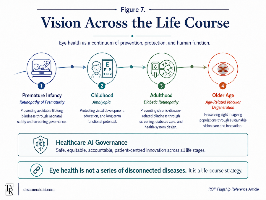
9.14 The ethical test for health-system leaders
For health-system leaders, the ethical question is not only whether an ROP policy exists. It is whether the system can look honestly at a premature infant and say: [47, 48, 49, 50, 51, 53]
We knew this baby was at risk.
We identified the baby in time.
We scheduled the first examination.
We completed follow-up.
We escalated uncertainty.
We arranged treatment when needed.
We protected discharge and transfer.
We informed and supported the parents.
We tracked the child until ROP surveillance was safely completed or care was formally transferred.
We did not leave vision to chance.
If a system cannot say this, then the issue is not only technical. It is ethical.
The child’s future should not depend on an accidental alignment of goodwill, memory, geography, technology, and parental persistence. It should depend on a designed pathway that recognises the child’s right to timely, competent, accountable care.
ROP prevention asks whether modern health systems can match the achievement of neonatal survival with the responsibility of lifelong vision protection. It asks whether survival will be accompanied by development, whether treatment will be accompanied by follow-up, whether technology will be accompanied by governance, and whether policy will be accompanied by performance.
The final message of Part IX is therefore clear: preventing ROP blindness is not only about saving sight. It is about protecting the child’s chance to participate fully in life.
Part IX has examined the equity, ethical, and lifelong consequences of preventable ROP blindness. The next section translates the article’s full argument into a practical policy brief for neonatal units, hospitals, ophthalmology departments, ministries, and regional networks.
Policy Brief: Preventing ROP Blindness through Neonatal Safety, Screening Governance, and Lifelong Vision Protection
Purpose of this policy brief
Retinopathy of Prematurity (ROP) is an important cause of childhood visual impairment and blindness in premature infants; much of this harm is preventable through timely screening, treatment and follow-up. [1, 3, 5, 14, 18, 19]
This policy brief translates the article’s core argument into practical actions for neonatal units, ophthalmology departments, hospital executives, ministries of health, regional networks, NGOs, child-eye-health programmes, and digital-health leaders.
The central policy message is simple:
Every premature infant at risk of ROP should be identified, entered into a screening pathway, examined at the right time, followed until risk is closed, treated urgently when needed, and supported through long-term vision care where indicated.
ROP blindness prevention requires more than a guideline. It requires a functioning pathway.
1. The policy problem
As neonatal care improves, more premature infants survive. This is a major achievement. However, survival must be accompanied by vision protection. When neonatal survival increases but ROP screening, treatment, and follow-up systems remain weak, health systems may unintentionally create a gap between survival and lifelong development. [1, 3, 5, 14, 35, 36]
This is the Survival–Vision Paradox: a child may survive prematurity but still lose vision because the system did not reliably connect neonatal care to eye screening, treatment, discharge safety, and follow-up.
ROP blindness may occur when:
- eligible infants are not identified;
- screening is not scheduled or occurs late;
- retinal findings are not graded accurately;
- treatment-requiring disease is not escalated urgently;
- treatment is unavailable or delayed;
- discharge or transfer interrupts follow-up;
- parents are not clearly informed;
- post-treatment surveillance is lost;
- long-term eye care is not arranged.
These failures are usually not the result of one person not caring. They are often pathway failures: gaps in responsibility, communication, data, workforce, equipment, referral, treatment access, and audit.
2. The leadership standard
A safe ROP programme should be able to answer “yes” to ten questions:
- Do we know which infants are eligible for ROP screening?
- Are all eligible infants entered into a live ROP register?
- Is the first examination date calculated and scheduled?
- Are repeat examinations tracked until risk is closed?
- Are findings documented clearly with zone, stage, vascular activity, follow-up interval, and next step?
- Is uncertain or severe disease escalated immediately?
- Can treatment-requiring ROP reach treatment within the required window?
- Is ROP status protected at discharge and transfer?
- Are parents informed, supported, and given clear written instructions?
- Do we audit delay, treatment access, follow-up loss, and outcomes?
If the answer to any of these is unclear, the programme is not yet safe enough.
3. Minimum safe-system requirements
Every health system caring for premature infants should establish the following minimum ROP safety architecture.
Table 9. Policy brief minimum safe-system requirements: essential ROP safety architecture
| Requirement | Why it matters |
| Clear eligibility criteria | Defines which infants must enter the pathway. |
| ROP register | Prevents eligible infants from becoming invisible. |
| First examination timing process | Converts eligibility into a scheduled action. |
| Repeat examination tracking | Ensures surveillance continues until risk is closed. |
| Trained examiners or imaging personnel | Protects screening quality and reliability. |
| Standardised documentation | Makes findings actionable and auditable. |
| Escalation pathway | Ensures severe or uncertain disease is reviewed urgently. |
| Treatment access or referral agreement | Connects detection to sight-saving intervention. |
| Discharge and transfer controls | Prevents pathway interruption across care transitions. |
| Parent communication and written information | Supports family understanding without shifting system responsibility onto parents. |
| Missed-visit tracking | Prevents follow-up loss from becoming invisible. |
| Audit and governance review | Turns policy into measurable safety performance. |
These are not luxury components. They are the basic architecture of a preventable-blindness pathway.
4. Priority actions for neonatal units
Neonatal units are the first link in the ROP prevention pathway.
Neonatal units should:
- identify all infants who meet local ROP screening criteria;
- enter eligible infants into a screening register;
- calculate the first examination date according to guideline timing;
- include ROP status in ward-round review for eligible infants;
- ensure oxygen governance, monitoring, equipment maintenance, and staff training;
- check ROP status before discharge or transfer;
- ensure parents know why screening matters and when the next appointment is due;
- confirm that outpatient follow-up is booked before discharge where needed;
- communicate retinal status clearly to receiving units during transfer;
- treat missed or overdue screening as a safety issue.
A neonatal unit should not rely on informal memory to trigger ROP screening. Eligibility should automatically lead to pathway entry. [1, 3, 5, 14]
5. Priority actions for ophthalmology departments
Ophthalmology departments are responsible for accurate retinal assessment, classification, escalation, treatment planning, and post-treatment follow-up. [6, 7, 1, 3, 5, 14]
Ophthalmology departments should:
- provide or arrange timely ROP screening for eligible infants;
- ensure examiners are trained and competent in ROP classification;
- document zone, stage, plus or pre-plus disease, aggressive features, image or view limitations, follow-up interval, urgency, and responsible clinician;
- escalate uncertain, severe, posterior, aggressive, or poor-quality cases;
- define when senior review, tele-ROP support, or referral is required;
- ensure treatment-requiring disease is not placed into routine referral pathways;
- maintain post-treatment surveillance, especially after anti-VEGF therapy;
- support long-term follow-up for refractive error, amblyopia, strabismus, and visual development;
- participate in multidisciplinary audit of missed, delayed, or lost follow-up cases.
The core ophthalmology standard is not only to examine the retina, but to ensure that every examination generates a safe next step.
6. Priority actions for hospital executives
Hospital leaders are responsible for the conditions that allow ROP care to be safe.
Hospital executives should:
- require a formal ROP pathway for every neonatal service caring for premature infants;
- ensure that the pathway includes eligibility capture, register, scheduling, examination, treatment access, discharge safety, transfer communication, and audit; [1, 3, 5, 14, 30, 31]
- appoint a named ROP lead or multidisciplinary ROP governance group;
- ensure sufficient workforce, equipment, space, imaging capacity, documentation systems, and referral agreements;
- define how treatment-requiring ROP will be managed before the first emergency occurs;
- ensure that ROP delays, missed examinations, treatment delays, and follow-up loss are reviewed as safety events;
- require regular reporting of ROP indicators to clinical governance or quality-safety committees;
- support parent information, interpreter access where needed, and written discharge instructions;
- ensure that digital tools, tele-ROP, or AI systems are introduced only within a governed pathway.
Hospital leadership should not ask, “Do we have ROP screening?”
It should ask, “Can we prove that every eligible infant is protected by the pathway?”
7. Priority actions for ministries of health and regional networks
ROP prevention requires regional and national coordination, especially where neonatal care is expanding or specialist ophthalmology capacity is limited. [1, 30, 31, 34, 51]
Ministries and regional networks should:
- include ROP prevention in neonatal safety, child-eye-health, public-health, and blindness-prevention strategies;
- define national or regional screening standards appropriate to local risk and neonatal-care conditions;
- map neonatal units, screening capacity, treatment centres, referral routes, and workforce gaps;
- ensure that neonatal expansion is matched by ROP screening and treatment capacity;
- create regional referral agreements for treatment-requiring ROP;
- support training for neonatal staff, ophthalmologists, nurses, imagers, and programme coordinators;
- develop tele-ROP models where geography or workforce limits access;
- monitor national or regional indicators for screening timeliness, treatment delay, follow-up loss, and outcomes;
- support equitable access for rural, remote, underserved, and resource-constrained communities;
- align AI and digital-health investment with validation, governance, treatment access, and human accountability.
A national ROP strategy should not only define who should be screened. It should define how the system will ensure that screening, treatment, and follow-up actually happen.
8. Priority actions for NGOs and child-eye-health programmes
NGOs and child-eye-health programmes can play an important role in supporting implementation, especially in resource-constrained settings. [18, 19, 20, 21, 22, 23]
They should:
- support neonatal-unit mapping and identification of underserved areas;
- help establish ROP registers and simple tracking systems;
- provide training support for screening, imaging, documentation, parent counselling, and follow-up coordination;
- strengthen referral pathways between district hospitals, neonatal units, and treatment centres;
- support procurement and maintenance of essential equipment where appropriate;
- help develop parent information materials in local languages;
- support tele-ROP or mobile screening models only when linked to treatment and follow-up;
- assist with audit, programme evaluation, and quality improvement;
- advocate for ROP prevention as part of national child-health and blindness-prevention strategies.
NGO support should strengthen local systems rather than create parallel pathways that disappear when external funding ends.
9. Priority actions for AI, tele-ROP, and digital-health programmes
AI and tele-ROP may extend screening capacity, but they must not be treated as substitutes for clinical governance.
Before implementing AI or tele-ROP, programmes should confirm:
- eligible infants are reliably identified;
- trained personnel can capture adequate images;
- image-quality standards are defined;
- images are reviewed within safe timeframes;
- AI performance is externally and locally validated;
- clinicians understand the tool’s intended use and limitations;
- positive, uncertain, or poor-quality results trigger escalation;
- treatment access or referral is available;
- parents are informed when AI-supported tools are used;
- data privacy, cybersecurity, consent, storage, and access are governed;
- false negatives, false positives, delay, and follow-up loss are audited;
- human accountability remains clearly assigned.
AI should strengthen the ROP pathway. It should not create false reassurance, weaken workforce planning, or generate alerts that no one acts upon. [41, 43, 44, 45, 46, 57]
In ROP, an algorithm does not prevent blindness. A governed pathway using the right tools at the right time prevents blindness.
10. The ROP Blindness Prevention Cascade for audit
Every programme should measure performance across the full prevention cascade:
Eligible infant → identified → registered → scheduled → screened on time → graded accurately → followed appropriately → treated within window if needed → monitored after treatment → safely discharged from ROP surveillance or transferred to long-term care [1, 3, 5, 14, 13, 47]
A practical ROP audit dashboard should include:
Table 10. ROP Blindness Prevention Cascade audit dashboard: minimum programme indicators
| Indicator | Minimum question |
| Eligibility capture | Were all eligible infants entered into the register? |
| First examination timing | Was the first examination completed within the guideline window? |
| Repeat examination reliability | Were follow-up examinations completed at safe intervals? |
| Documentation completeness | Did each record include findings, urgency, follow-up interval, and next step? |
| Grading quality | Were uncertain or poor-quality cases escalated? |
| Treatment escalation | How long from treatment-requiring diagnosis to treatment decision? |
| Treatment delivery | How long from treatment decision to treatment completion? |
| Discharge safety | Was ROP status and follow-up plan documented before discharge? |
| Transfer safety | Was ROP status accepted by the receiving unit? |
| Post-treatment surveillance | Were treated infants followed until risk was closed? |
| Follow-up loss | How many infants were lost before safe discharge from surveillance? |
| Parent communication | Did families receive verbal and written information? |
The purpose of audit is not bureaucracy. It is to detect weakness before a child is harmed.
11. Red flags requiring urgent programme review
A neonatal or regional ROP programme should trigger immediate review if any of the following occur:
- infants are discharged without documented ROP status;
- no live register exists;
- first examinations are missed or regularly delayed;
- follow-up intervals are not tracked;
- treatment-requiring disease is escalated through routine referral systems;
- no treatment pathway or referral agreement exists;
- anti-VEGF treatment is used without prolonged surveillance tracking;
- transfers occur without retinal status handover;
- families report unclear instructions or missing appointment information;
- missed ROP appointments do not trigger active tracing;
- tele-ROP images are captured but not reviewed within safe timeframes;
- AI or digital tools are introduced without validation, governance, and treatment access;
- audit data are collected but not reviewed by accountable leaders.
These red flags indicate that the programme may exist in name but not yet in dependable practice.
12. Implementation priorities: first 100 days
For a hospital or regional network beginning or strengthening an ROP programme, the first 100 days should focus on reliability before sophistication. [30, 31, 32, 33, 34]
Priority actions:
- Appoint a named ROP lead or multidisciplinary ROP governance group.
- Confirm local eligibility criteria and first-examination timing rules.
- Create or update a live ROP register.
- Identify all currently eligible infants in neonatal care.
- Establish a weekly due-list review.
- Standardise ROP documentation templates.
- Define escalation rules for severe, uncertain, posterior, aggressive, or poor-quality cases.
- Confirm where treatment can be delivered and how urgent referral will occur.
- Add ROP status to discharge and transfer checklists.
- Create parent information materials and contact routes.
- Begin basic audit of eligibility capture, first examination timing, treatment delay, and follow-up loss.
- Review all overdue or missed cases as safety events.
The first goal is not perfection. The first goal is that no eligible infant is invisible.
13. Twelve-month priorities
Within 12 months, programmes should aim to move from basic pathway existence to measurable performance.
Priorities:
- audit all eligible infants across the full prevention cascade;
- review treatment-requiring cases and treatment delay intervals;
- strengthen regional referral agreements;
- train additional examiners, imagers, nurses, and coordinators;
- review oxygen governance and neonatal safety processes;
- improve parent communication and discharge materials;
- reduce follow-up loss through active tracing;
- review outcomes after laser and anti-VEGF treatment;
- establish regular reporting to hospital or regional governance bodies;
- assess whether tele-ROP or digital tools are needed and safe to introduce.
A 12-month review should answer: is the programme more reliable than it was at baseline?
14. Longer-term system priorities
Over three to five years, mature ROP systems should aim to:
- integrate neonatal and ophthalmology data systems;
- develop regional or national ROP dashboards;
- expand workforce training and certification;
- support equitable access for rural and remote neonatal units;
- create sustainable treatment networks;
- evaluate tele-ROP and AI tools through governed implementation;
- link ROP outcomes to neonatal quality improvement;
- connect paediatric ophthalmology follow-up with child-development and education support;
- reduce avoidable ROP blindness and severe visual impairment;
- build ROP prevention into life-course eye-health strategies.
Where feasible, regional or national ROP outcomes registries should also be developed to capture eligibility, screening timing, disease severity, treatment, retreatment, follow-up loss and long-term visual outcomes. Such registries should support benchmarking, quality improvement and responsible research, while protecting privacy, consent, data security and appropriate governance. [12, 13, 24, 25, 33, 34]
Long-term success should not be measured only by screening activity. It should be measured by whether eligible infants are protected from preventable lifelong visual loss. [1, 3, 5, 14, 13, 47]
15. Policy recommendations
Recommendation 1: Treat ROP as a neonatal safety priority
ROP prevention should be embedded into neonatal quality systems, not treated as an optional ophthalmology referral.
Recommendation 2: Require a live ROP register
Every neonatal unit caring for premature infants should maintain a reliable register of eligible infants, due dates, findings, treatment status, discharge status, and follow-up outcomes. [1, 3, 4]
Recommendation 3: Make discharge and transfer ROP-safe
No eligible premature infant should be discharged or transferred without documented ROP status, next examination date, responsible service, and parent information. [1, 3, 5, 14]
Recommendation 4: Link screening to treatment access
Screening programmes must have defined treatment pathways, referral agreements, transfer processes, and post-treatment surveillance. [1, 3, 5, 14]
Recommendation 5: Audit the full pathway
Audit should measure eligibility capture, examination timing, documentation, escalation, treatment delay, discharge safety, transfer safety, parent communication, and follow-up loss. [1, 3, 30, 31, 34]
Recommendation 6: Support parents without blaming them
Parents should receive clear information, written instructions, confirmed appointments, and contact routes. They should not be expected to compensate for system gaps. [1, 30, 31, 34, 51]
Recommendation 7: Govern AI and tele-ROP responsibly
Digital innovation should be clinically validated, ethically governed, human-supervised, linked to treatment access, and monitored for equity and safety. [41, 43, 44, 45, 46, 57]
Recommendation 8: Connect ROP prevention to life-course vision protection
ROP should be recognised as the neonatal pillar of a wider life-course vision strategy, alongside amblyopia, diabetic retinopathy, age-related macular degeneration, and healthcare AI governance.
16. The final policy message
ROP prevention is a test of whether a health system can convert knowledge into protection.
Guidelines are necessary, but insufficient.
Equipment is necessary, but insufficient.
Technology is necessary, but insufficient.
Professional commitment is necessary, but insufficient. [1, 30, 31, 34, 51]
What protects the infant is the complete pathway: eligibility recognition, register entry, timed screening, accurate grading, urgent escalation, treatment access, discharge safety, parent support, post-treatment surveillance, audit, and long-term vision follow-up.
A premature infant should not depend on chance, geography, memory, parental persistence, or institutional goodwill to receive sight-saving care.
The policy goal should be clear:
No eligible infant invisible.
No screening window missed.
No severe disease left without action.
No discharge without retinal status.
No treatment without surveillance.
No child left to carry preventable Lifelong Vision Debt.
Preventing ROP blindness is not only about saving sight. It is about completing the promise of neonatal survival with lifelong vision protection.
The intended contribution of this article is therefore translational as well as scholarly: to help clinicians, neonatal teams, hospital leaders, policymakers and AI implementers turn established ROP evidence into programme design, audit, accountability and implementation practice.
The named frameworks developed in this article are offered as citable conceptual tools for programme design, audit, policy dialogue and translational implementation, while clinical decisions must continue to follow recognised ROP guidance, local protocols and specialist judgement.
Parent and Professional FAQs
These FAQs are designed to support clarity, trust, and practical understanding. They are educational and should not replace advice from the baby’s neonatal, ophthalmology, or paediatric care team.
A. Parent FAQs
1. What is Retinopathy of Prematurity?
Retinopathy of Prematurity, often called ROP, is an eye condition that can affect babies born very early or with very low birth weight. It happens because the blood vessels at the back of the eye, in a layer called the retina, may still be developing after premature birth. In some babies, this blood-vessel growth can become abnormal. [1, 2, 3, 6, 7, 9]
ROP can be mild and improve on its own, but in some babies it can become serious. This is why timely eye screening is important.
2. Why does my premature baby need eye screening?
Premature babies may need eye screening because ROP can develop silently. A baby may look well, feed well, and still have changes developing inside the eye that parents cannot see. [1, 3, 5, 14]
Screening allows the ophthalmology team to check whether the retina is developing safely. The goal is to detect concerning changes early, before they threaten vision.
3. Does every premature baby need ROP screening?
Not every premature baby needs ROP screening. Screening is usually recommended for babies who were born very early, had very low birth weight, or had a neonatal course that increases risk. The exact criteria vary by country, hospital, and clinical guideline. [1, 3, 5, 14]
Parents should feel able to ask the neonatal team: “Does my baby meet the criteria for ROP screening, and when is the first eye examination due?”
4. Is ROP my fault?
No. ROP is not the fault of parents. It is not caused by anything a mother or father did or failed to do.
ROP is linked to prematurity, immature retinal development, neonatal illness, oxygen exposure, growth factors, and the complex medical needs of very small or very early babies. Oxygen can be lifesaving in neonatal care, but it also needs careful monitoring because the immature retina is sensitive during this stage of development. [16, 17, 18, 6, 7]
5. What happens during an ROP eye examination?
The baby may be given eye drops to make the pupils larger so that the retina can be examined properly. The eye examination may be performed directly by an ophthalmologist or trained specialist team. In some hospitals, retinal photographs may be taken using specialised wide-field imaging equipment. [1, 3, 5, 14, 35, 36]
The examination can be uncomfortable for a short time, but it is done to protect the baby’s long-term vision.
6. Does ROP always need treatment?
No. Many babies who are screened for ROP do not develop severe disease and do not need treatment. Some babies develop mild ROP that improves on its own with careful monitoring. [1, 2, 3, 6, 7, 9]
A smaller number develop more serious ROP that may need treatment to reduce the risk of retinal detachment and permanent visual loss. The decision depends on the severity, location, and behaviour of the disease.
7. What treatments are used for serious ROP?
The main treatments for serious ROP include laser treatment and carefully administered anti-VEGF injection therapy. In more advanced cases, where retinal detachment has developed, surgery may be considered. [9, 10, 11, 26, 27, 28]
The specialist team should explain what has been found, why treatment is recommended, what the treatment involves, what risks and alternatives exist, and what follow-up will be needed afterwards.
8. Why are repeat appointments needed?
ROP can change over time. It may improve, stay stable, worsen, recur, or reactivate after treatment. This means that one eye examination is often not enough. [1, 3, 5, 14]
Repeat appointments are not ordinary routine appointments. They are part of a time-sensitive safety pathway. Missing or delaying follow-up can mean that important changes are not detected at the right time.
9. Why does follow-up still matter after treatment?
Follow-up after treatment is essential. The team needs to check whether the ROP is improving, whether more treatment is needed, and whether there is any recurrence or reactivation. [9, 10, 11, 26, 27, 28]
Follow-up is especially important after anti-VEGF injection therapy because disease activity may return later. Later childhood eye care may also be needed to check for glasses, squint, amblyopia, or other visual-development concerns.
10. What should I ask before my baby leaves the neonatal unit?
Before discharge or transfer, parents can ask:
- Does my baby need ROP screening?
- Has the first eye examination been done?
- What were the findings?
- Is another eye examination needed?
- When and where is the next appointment?
- Who is responsible for arranging the appointment?
- Who should I contact if the appointment letter does not arrive?
- What happens if my baby is transferred to another hospital?
- Does the discharge summary clearly state the ROP plan?
These questions do not mean parents are responsible for carrying the pathway alone. They help make the plan visible.
11. What should I do if the appointment letter does not arrive?
Do not assume that no appointment means no follow-up is needed. Contact the neonatal unit, ophthalmology department, paediatric team, or the contact number you were given. [1, 3, 5, 14]
ROP follow-up can be time-sensitive. It is safer to ask early than to wait and risk missing the correct examination window.
12. Should I be frightened if my baby needs ROP screening?
The need for screening does not mean that your baby will lose vision. Many babies who are screened do not need treatment. Screening is done because early detection and careful follow-up can protect sight. [1, 3, 5, 14]
The message should be realistic but reassuring: ROP can be serious, but it is also one of the clearest examples of a condition where timely screening, careful monitoring, and appropriate treatment can protect vision.
13. What is the role of parents in the ROP pathway?
Parents are essential partners. They can ask questions, attend appointments, keep contact details updated, and tell the team if an appointment is missing or unclear. [35, 36, 37, 38, 39, 40]
However, parents should not be made to feel that they are carrying the system alone. A safe ROP pathway should include a clear plan, a documented appointment, a responsible team, and a reliable follow-up system.
14. What is the most important message for families?
A baby should not depend on memory, chance, or parental persistence alone to receive sight-saving screening. The safest pathway is one in which parents, neonatal teams, ophthalmology teams, and hospital systems work together. [1, 3, 5, 14]
The goal is not to frighten families. The goal is to make sure that every premature baby who needs eye screening is identified, examined, followed, and treated at the right time.
B. Professional and Leadership FAQs
1. Is ROP mainly an ophthalmology problem?
No. ROP is a retinal disease, but ROP blindness prevention is a neonatal safety and health-system governance pathway. Ophthalmology is essential, but safe prevention also depends on neonatal teams, nursing, oxygen governance, registers, discharge planning, transfer communication, treatment access, parent information, and audit. [16, 17, 18, 1, 3, 5]
ROP should be treated as a shared safety pathway, not as an optional ophthalmology referral.
2. What is the minimum safe ROP system?
A minimum safe ROP system requires clear eligibility criteria, a live register, first examination timing, repeat examination tracking, trained examination or imaging capacity, standardised documentation, escalation rules, treatment access or referral agreements, discharge and transfer checks, parent information, missed-visit tracking, and audit. [1, 3, 5, 14, 30, 31]
A programme may begin simply, but it must begin with reliability.
3. Why is a live ROP register so important?
The register prevents eligible infants from becoming invisible. It should show who is eligible, who is due, who is overdue, who has been examined, what the findings were, when follow-up is due, who needs treatment, who has been transferred, who has been discharged, and who remains under surveillance. [1, 3, 4]
A register should not be a passive list. It should generate action.
4. What is the Third Delay in ROP Blindness?
The Third Delay in ROP Blindness framework analyses preventable ROP blindness through three predictable delay domains:
- Eligibility recognition delay — the at-risk infant is not identified, registered, or tracked safely.
- Screening and grading delay — the examination is missed, late, unavailable, inadequately documented, or not interpreted and escalated safely. [1, 3, 5, 14, 30, 31]
- Treatment and follow-up delay — the infant is diagnosed, but treatment, transfer, consent, parent communication, specialist availability, or post-treatment surveillance fails.
The framework is not intended to blame individuals. It is designed to identify where the pathway failed and how the system can be redesigned.
5. Should screening and grading delay be separated?
Operationally, yes. Within the Third Delay framework, Delay 2 deliberately contains two related but distinct subdomains: examination timing and grading or decision quality. [30, 31, 32, 33, 34, 35]
An infant may be harmed because the examination is missed, late, or unavailable. An infant may also be harmed because the examination occurs, but the findings are not interpreted, documented, or escalated with sufficient accuracy. The three-delay model remains useful and memorable, while programme audit should still separate screening timeliness from grading and decision quality.
6. What should happen when findings are uncertain?
Uncertainty should trigger protection, not passive delay.
Poor retinal view, poor image quality, suspected plus disease, possible aggressive ROP, posterior disease, unclear zone, disagreement between graders, or incomplete documentation should trigger senior review, repeat examination, tele-ROP support, referral, or escalation according to risk. [6, 7, 35, 36, 37, 38]
Uncertainty is not failure if it is recognised and escalated. It becomes dangerous when it is hidden inside vague documentation or treated as reassurance.
7. What should be documented after every ROP examination?
Every ROP examination should document findings in a way that creates a safe next step. This should include, where applicable: [1, 2, 3, 6, 7, 9]
- retinal zone;
- disease stage;
- plus or pre-plus disease;
- aggressive features;
- image or view limitations;
- whether treatment is required;
- recommended follow-up interval;
- urgency;
- responsible clinician or service;
- parent communication where relevant.
A screening note without a follow-up plan is an incomplete safety document.
8. How should discharge and transfer be made ROP-safe?
Discharge and transfer should trigger retinal safety checks.
Before discharge or transfer, the team should confirm eligibility, last examination findings, next examination date, treatment status, whether follow-up is still required, who is responsible, whether parents have received written information, and whether the receiving team has accepted responsibility. [1, 3, 5, 14]
“Outpatient follow-up” is not enough unless the date, service, contact route, and urgency are clear.
9. What is the main leadership test for an ROP programme?
The leadership test is not simply, “Do we have ROP screening?”
The better question is: “Can we prove that every eligible infant moves safely through the full pathway?”
Leaders should be able to see the ROP register, overdue list, first examination timing, treatment delay intervals, discharge safety process, transfer handover process, follow-up loss rate, parent communication process, equipment status, workforce plan, and audit record. [1, 3, 5, 14, 30, 31]
10. How should treatment access be built into the pathway?
Treatment access should be planned before severe disease is diagnosed. The pathway should define where treatment can occur, who provides it, how urgent contact is made, how parents are counselled, how neonatal or anaesthetic support is arranged, how transfer occurs if needed, and how post-treatment surveillance is tracked. [1, 3, 5, 14, 12, 13]
Screening without a reliable treatment pathway is a dangerous half-system.
11. What is the role of oxygen governance in ROP prevention?
Oxygen governance is part of upstream ROP prevention. Oxygen can be lifesaving for premature infants, but it must be carefully monitored because oxygen exposure and fluctuation can influence the immature retina. [16, 17, 18, 30, 31, 32]
Oxygen governance means appropriate targets, monitoring, alarm response, equipment maintenance, staff training, documentation, and review of practice. It should be framed as neonatal safety, not as blame.
12. How should anti-VEGF treatment be governed?
Anti-VEGF therapy may be valuable in selected infants, especially in posterior, aggressive, or medically complex disease. However, it requires informed consent, specialist judgement, local protocol, documentation, and prolonged surveillance because recurrence or reactivation may occur later. [9, 10, 11, 26, 27, 28]
A programme should not use anti-VEGF therapy unless it can provide the follow-up that the treatment requires.
13. Can tele-ROP or AI replace ophthalmologists?
No. Tele-ROP and AI may extend capacity, support triage, assist image review, improve consistency, and help programme audit, but they do not replace clinical accountability. [30, 31, 32, 33, 34, 35]
AI cannot identify infants who were never registered. It cannot act on reports that no one reviews. It cannot provide treatment access where none exists. It cannot counsel parents or carry professional responsibility for a child’s sight.
In ROP, an algorithm does not prevent blindness. A governed pathway using the right tools at the right time prevents blindness.
14. What governance should exist before AI is introduced?
Before AI is introduced, a programme should have reliable eligibility capture, a functioning register, trained imaging or examination personnel, image-quality standards, secure data systems, validated AI performance for the intended use, local performance review, human oversight, override rules, escalation pathways, treatment access, parent communication, data governance, and audit. [1, 3, 5, 14, 30, 31]
AI should strengthen a pathway. It should not be used to compensate for missing basics.
15. How should programmes measure ROP safety?
ROP safety should be measured across the full prevention cascade:
Eligible infant → identified → registered → scheduled → screened on time → graded accurately → followed appropriately → treated within window if needed → monitored after treatment → safely discharged from ROP surveillance or transferred to long-term care [1, 3, 5, 14, 13, 47]
Useful indicators include eligibility capture, first examination timing, repeat examination reliability, documentation completeness, grading quality, treatment escalation time, treatment delivery time, discharge safety, transfer safety, post-treatment surveillance, follow-up loss, and parent communication.
16. What should trigger urgent programme review?
Urgent review is needed when infants are discharged without ROP status, no live register exists, first examinations are missed or delayed, follow-up intervals are not tracked, treatment-requiring disease is escalated routinely rather than urgently, no treatment route exists, transfers occur without retinal handover, anti-VEGF is used without prolonged follow-up tracking, tele-ROP reports are delayed, AI tools are introduced without governance, or audit data are collected but not acted upon. [9, 10, 11, 26, 27, 28]
These are not minor administrative issues. They are safety signals.
17. How should missed appointments be handled?
Missed ROP follow-up should trigger active tracing and pathway review. The programme should ask whether the appointment was booked, whether parents were informed, whether contact details were correct, whether transport or cost barriers existed, whether reminders were sent, and whether the missed visit was escalated. [1, 3, 5, 14, 35, 36]
A missed ROP appointment should not be treated as ordinary outpatient non-attendance. The disease may progress silently while the baby appears well.
18. What is Lifelong Vision Debt?
Lifelong Vision Debt describes the long-term burden created when a preventable neonatal vision-protection opportunity is missed or incompletely followed through. [18, 19, 20, 21, 22, 23]
A missed screening, delayed treatment, unsafe transfer, or lost follow-up may create consequences that extend into childhood development, education, family wellbeing, social participation, and adult independence.
The phrase is intended to remind health systems that the cost of missed prevention does not disappear. It is transferred forward to the child, family, and society.
19. What is the most important message for policymakers?
ROP prevention should be embedded into neonatal safety, child-eye-health, and public-health strategy. It should not be treated as a specialist issue that begins and ends in the eye clinic. [18, 19, 20, 21, 22, 23]
The policy test is whether every eligible infant has a reliable route from risk recognition to screening, treatment, follow-up, and long-term vision protection.
20. What is the final practical message for professionals and leaders?
Guidelines are necessary, but insufficient. Equipment is necessary, but insufficient. Technology is necessary, but insufficient. Professional commitment is necessary, but insufficient. [1, 2, 3, 6, 7, 9]
What protects the infant is the complete pathway: eligibility recognition, register entry, timed screening, accurate grading, urgent escalation, treatment access, discharge safety, parent support, post-treatment surveillance, audit, and long-term vision follow-up.
No eligible infant should be invisible. No screening window should be missed. No severe disease should be left without action. No treatment should occur without surveillance. No child should be left to carry preventable Lifelong Vision Debt.
Definitive Glossary of ROP and Neonatal Safety Terminology
This glossary is organised by function rather than alphabetically so that clinical, pathway, governance, AI, and public-health concepts can be followed easily. It is designed as a practical reading aid for clinicians, neonatal teams, hospital leaders, policymakers, and digital-health stakeholders.
Definitions are intentionally concise. They explain how each term is used in this article and how it relates to ROP blindness prevention, neonatal safety, screening governance, treatment access, and lifelong vision protection.
A1. Prematurity, retinal development, and core ROP terms
Table 11. Glossary A1: prematurity, retinal development, and core ROP terms
| Term | Meaning in this article |
| Retinopathy of Prematurity (ROP) | Retinopathy of Prematurity is abnormal retinal vascular development in premature infants. It ranges from mild self-resolving disease to severe sight-threatening disease requiring urgent treatment and disciplined follow-up. |
| Prematurity | Prematurity refers to birth before full-term gestation and is the biological starting point for ROP risk because retinal vascular development may still be incomplete at the time of birth. |
| Birth weight | Birth weight is the infant’s weight at birth and is one of the key criteria used with gestational age and clinical risk to determine ROP screening eligibility. |
| Gestational age | Gestational age is the length of pregnancy at the time of birth, usually measured in weeks, and is a major determinant of prematurity-related ROP risk and screening eligibility. |
| Postmenstrual age | Postmenstrual age is the infant’s gestational age at birth plus chronological age after birth, and it is used to time the first and subsequent ROP examinations. |
| Retina | The retina is the light-sensitive tissue at the back of the eye, and in premature infants its blood-vessel development may be incomplete, creating vulnerability to ROP. |
| Retinal vascularisation | Retinal vascularisation is the development of blood vessels across the retina, a process that may be incomplete in premature infants and may become abnormal in ROP. |
A2. ROP classification, activity, and severity terms
Table 12. Glossary A2: ROP classification, activity, and severity terms
| Term | Meaning in this article |
| Zone | Zone describes the location of ROP within the retina, with more posterior disease generally carrying higher risk and requiring closer attention. |
| Stage | Stage describes the severity of ROP changes at the border between vascularised and avascular retina, ranging from mild demarcation to advanced retinal detachment. |
| Plus disease | Plus disease describes abnormal dilation and tortuosity of posterior retinal blood vessels and is a key marker of active, more severe ROP that may indicate the need for urgent treatment. |
| Pre-plus disease | Pre-plus disease describes retinal vascular abnormality that is more concerning than normal but not severe enough to meet the definition of plus disease, usually requiring closer follow-up and careful clinical judgement. |
| Aggressive ROP | Aggressive Retinopathy of Prematurity is a rapidly progressive form of ROP that may develop severe vascular activity and sight-threatening progression quickly, requiring urgent recognition, senior review, and timely treatment planning. |
| Type 1 ROP | Type 1 ROP is treatment-requiring disease defined as: any stage of ROP in Zone I with plus disease; stage 3 ROP in Zone I without plus disease; or stage 2 or 3 ROP in Zone II with plus disease. |
| Treatment-requiring ROP | Treatment-requiring ROP refers to disease that has reached a level of severity, location, vascular activity, or progression where intervention is needed to reduce the risk of retinal detachment and permanent visual loss. |
| Retinal detachment | Retinal detachment is a serious complication in which the retina separates from its normal position, and in advanced ROP it may cause severe permanent visual loss or blindness. |
B. Treatment, recurrence, and long-term vision protection
Table 13. Glossary B: treatment, recurrence, and long-term vision protection
| Term | Meaning in this article |
| Laser photocoagulation | Laser photocoagulation is an established ROP treatment that applies laser to peripheral avascular retina to reduce the drive for abnormal vessel growth and prevent progression to retinal detachment. |
| Anti-VEGF therapy | Anti-VEGF therapy is an intravitreal treatment that suppresses vascular endothelial growth factor activity in selected cases of treatment-requiring ROP. It requires strict governance and prolonged follow-up because late disease reactivation may occur. |
| Vascular endothelial growth factor (VEGF) | Vascular endothelial growth factor is a biological signal involved in blood-vessel growth, and abnormal VEGF-driven activity is one reason anti-VEGF therapy may be used in selected cases of serious ROP. |
| Recurrence | Recurrence refers to the return of active ROP after apparent improvement or response to treatment, requiring renewed clinical assessment and possible retreatment. |
| Reactivation | Reactivation refers to renewed ROP activity after apparent regression, particularly important after anti-VEGF therapy because disease activity may return later and requires prolonged surveillance. |
| Long-term paediatric ophthalmology follow-up | Long-term paediatric ophthalmology follow-up is continued eye care after acute ROP has regressed or been treated, focusing on refractive error, amblyopia, strabismus, retinal sequelae, visual development, and functional vision. |
| Amblyopia | Amblyopia is reduced vision caused by abnormal visual development in childhood, and it may occur after ROP if refractive error, strabismus, or visual deprivation is not identified and managed appropriately. |
| Strabismus | Strabismus, often called squint, is misalignment of the eyes and may occur in children born prematurely or affected by ROP, requiring assessment because it can affect binocular vision and amblyopia risk. |
| Functional vision protection | Functional vision protection means preserving not only retinal anatomy, but also the child’s ability to use vision for development, learning, mobility, communication, independence, play, and participation. |
C1. Screening eligibility, follow-up, and register terminology
Table 14. Glossary C1: screening eligibility, follow-up, and register terminology
| Term | Meaning in this article |
| Eligibility criteria | Eligibility criteria are the local or national rules used to identify which premature infants require ROP screening, usually based on gestational age, birth weight and, in some settings, additional clinical risk factors. Postmenstrual and chronological age are then used, with local guidance, to determine when screening should begin. |
| Follow-up interval | Follow-up interval is the recommended time between ROP examinations, determined by retinal findings, disease activity, infant age, prior treatment, and local guideline requirements. |
| Follow-up loss | Follow-up loss occurs when an infant who still needs ROP surveillance does not complete the required examinations, treatment review, or long-term eye-care pathway before risk has been safely closed. |
| ROP register | An ROP register is a live tracking system that records eligible infants, examination due dates, findings, follow-up intervals, treatment status, transfer status, discharge plans, and outcomes so that no at-risk infant becomes invisible. |
C2. Governance, transfer, audit, and oxygen-safety terminology
Table 15. Glossary C2: governance, transfer, audit, and oxygen-safety terminology
| Term | Meaning in this article |
| Screening governance | Screening governance is the system of responsibilities, registers, scheduling controls, documentation standards, escalation rules, audit indicators, and leadership oversight that ensures ROP screening happens safely and reliably. |
| Discharge safety | Discharge safety means that a premature infant leaves the neonatal unit only with clear documentation of ROP status, next appointment, responsible service, parent information, and a plan for ongoing surveillance where needed. |
| Neonatal transfer pathway | The neonatal transfer pathway is the process by which an infant moves between units or hospitals, and it must include clear communication of ROP eligibility, screening status, last findings, treatment status, next due date, and responsible receiving service. |
| Referral pathway | A referral pathway is the defined route by which infants with severe, uncertain, or treatment-requiring ROP are escalated to senior review, specialist treatment, transfer, or regional expert support. |
| Clinical accountability | Clinical accountability means that named professionals or services remain responsible for interpreting findings, making decisions, escalating risk, communicating with parents, arranging treatment, and ensuring follow-up is completed. |
| Audit | Audit is the structured review of programme performance, including eligibility capture, screening timing, documentation, treatment delay, discharge safety, transfer safety, and follow-up loss, to identify weaknesses before children are harmed. |
| Oxygen governance | Oxygen governance refers to the safe use, monitoring, documentation, equipment maintenance, alarm response, staff training, and audit of oxygen therapy in premature infants, recognising that oxygen can be lifesaving while requiring careful control. |
D. Author-developed and adapted conceptual frameworks
Table 16. Glossary D: Author-developed and adapted conceptual Frameworks
The following entries define the author-developed and adapted conceptual frameworks introduced in this article. Some are original synthesis constructs; others adapt established public-health or patient-safety concepts to the ROP pathway. They are provided to support consistent interpretation, citation and programme discussion and should be applied in conjunction with local clinical protocols, national guidance and specialist judgement.
| Term | Meaning in this article |
| NICU-to-Retina Safety Chain | The NICU-to-Retina Safety Chain is introduced in this article as a systems-level construct describing the full pathway from neonatal survival and oxygen governance to eligibility recognition, retinal screening, disease grading, treatment access, discharge safety, follow-up and long-term vision protection. |
| ROP Blindness Prevention Cascade | The ROP Blindness Prevention Cascade is proposed in this article as a measurable pathway through which an eligible infant is identified, registered, scheduled, screened, graded, followed, treated when needed, monitored after treatment, and safely discharged from surveillance or transferred to long-term care. |
| Third Delay in ROP Blindness | An author-developed adaptation of established delay frameworks to the ROP pathway, comprising delays in eligibility recognition, screening and grading, and treatment or follow-up. |
| Survival–Vision Paradox | A conceptual framing describing the risk that improved neonatal survival may be accompanied by a growing burden of avoidable visual impairment when screening, treatment and follow-up capacity do not expand proportionately. |
| Lifelong Vision Debt | Lifelong Vision Debt is introduced in this article to describe the long-term developmental, educational, social, emotional, family and economic burden created when a preventable neonatal vision-protection opportunity is missed or incompletely followed through. |
| ROP programme maturity | ROP programme maturity describes how reliably a service moves from basic screening availability towards a governed, audited, treatment-linked and equitable programme. AI may be incorporated where appropriate but is not required for programme maturity. |
| Vision Across the Life Course | Vision Across the Life Course is a public-health framing that links ROP in premature infancy, amblyopia in childhood, diabetic retinopathy in adulthood, age-related macular degeneration in older age, and healthcare AI governance across all stages. This is a public-health framing, not a disease-progression model. |
These conceptual synthesis tools are not clinical prediction rules, screening criteria, treatment algorithms or substitutes for locally approved clinical guidance.
E. Digital health, tele-ROP, and AI governance terms
Table 17. Glossary E: digital health, tele-ROP, and AI governance terms
| Term | Meaning in this article |
| Wide-field retinal imaging | Wide-field retinal imaging captures broad retinal photographs in premature infants and can support tele-ROP, documentation, expert review, AI-assisted analysis, quality assurance, and longitudinal comparison. |
| Tele-ROP | Tele-ROP is an image-based screening model in which retinal images are captured locally and reviewed remotely by qualified experts, extending specialist reach when supported by reliable imaging, reporting, escalation, and treatment pathways. |
| AI-supported screening | AI-supported screening refers to the use of artificial intelligence to assist image assessment, triage, severity scoring, quality control, or audit. Clinical responsibility remains with trained professionals and accountable services. |
| Data governance | Data governance is the safe, ethical, secure, and accountable handling of clinical data, retinal images, AI outputs, audit records, privacy, consent, storage, access, and cybersecurity. |
| Human oversight | Human oversight means that trained clinicians or accountable teams review, interpret, confirm, override, escalate, and act on screening findings or AI outputs rather than allowing technology to function as an autonomous safety replacement. |
F. Public-health and equity terms
Table 18. Glossary F: public-health and equity terms
| Term | Meaning in this article |
| Childhood blindness | Childhood blindness refers to severe visual impairment or loss of sight occurring in childhood, and preventable ROP blindness is especially important because early-life visual loss can affect development, education, family wellbeing, and life-course opportunity. |
Note: This glossary supports interpretation of the article. It does not replace local clinical guidelines, neonatal-unit protocols, specialist ophthalmology judgement, or urgent clinical review for an individual infant.
Back Matter
Methods and Publication Transparency
This section describes the article’s evidence base and methodology and provides publication-transparency information concerning authorship, AI-assisted editorial use, publication status, competing interests, funding, data availability, ethics, licensing, version control, and future updates. These statements are provided to support transparency, accountability, responsible interpretation, and appropriate citation of this work.
Methods and evidence statement
This article presents an evidence-informed narrative and policy analysis rather than a systematic review or clinical practice guideline. It brings together established clinical evidence, international screening and treatment guidance, public-health and patient-safety principles, health-system and digital-health governance, and child-rights perspectives relevant to Retinopathy of Prematurity (ROP).
Sources were identified iteratively through targeted searches of clinical guideline repositories, professional and governmental websites, and relevant published literature, supplemented by reference-list review. The evidence base was reviewed and updated up to 18 July 2026. Priority was given to current national and international guidance, consensus classification statements, landmark clinical trials, systematic reviews, population-surveillance studies, and key literature on public health, patient safety and digital-health governance. Source selection was purposive and relevance-led; this was not a systematic review, and no protocol-driven comprehensive search, formal risk-of-bias assessment or meta-analysis was undertaken.
Particular attention is given to guidance and classification documents from the United Kingdom, United States and Australasia, alongside international ROP terminology and evidence on treatment-requiring disease. The analysis also draws on tele-ROP and AI-supported screening literature, Indian programme experience, and wider eye-health and disability-rights frameworks.
The analysis uses a critical interpretive synthesis of the international ROP literature to develop a systems-level governance framework. Where existing terminology did not adequately capture important relationships across the care pathway, the NICU-to-Retina Safety Chain, the ROP Blindness Prevention Cascade, the Survival–Vision Paradox and Lifelong Vision Debt were developed as author synthesis constructs. The Third Delay in ROP Blindness is an author-developed adaptation of the established Three Delays model from maternal-health analysis to the ROP pathway. [32]
These constructs are intended to support clinical reflection, programme design, policy discussion, audit and health-system strengthening. They have not been externally validated as composite clinical or programme tools and do not replace locally approved screening criteria, treatment protocols, specialist judgement or urgent review.
No new patient-level data were collected or analysed. The article does not report a clinical trial and includes no individual patient images or identifiable clinical information.
Important evidence gaps remain. These include the optimal duration and intensity of surveillance after anti-VEGF therapy; safe and efficient screening intervals across settings with differing disease prevalence and workforce capacity; and long-term visual, refractive and neurodevelopmental outcomes following different treatment and follow-up pathways. These uncertainties should remain explicit, but they should not delay implementation of established pathway principles. Instead, they should inform continuous audit, active surveillance, registry-based learning and responsible research. [9, 10, 11, 14, 26, 27, 28, 29, 47, 48, 49, 50]
AI-use disclosure
Generative AI tools were used during the development of this article to support language refinement, structural organisation, consistency checking, and editorial review. Dr. Samer Al-Diri conceived the article, selected, interpreted and reviewed the evidence, developed the clinical and health-systems arguments and named conceptual frameworks, and accepts full responsibility for the final content. AI tools were not used to generate or analyse patient-level data, replace source verification, or make clinical decisions.
AI-assisted design tools were used to support the visual rendering of author-developed concepts. Dr. Samer Al-Diri reviewed and approved the content.
Author
Ophthalmologist and Health-Systems Governance Researcher
Professional profiles: ORCID iD: 0009-0004-1908-0714 | ResearchGate: Samer Al-Diri | Website: drsameraldiri.com
Dr. Samer Al-Diri is a UK-trained ophthalmologist with postgraduate expertise in retina, public health, healthcare management, and health-system transformation. His work focuses on ophthalmology, preventable blindness, public health, healthcare AI governance, digital health, patient-centred accountability, and resilient health-system design.
This article forms part of Dr. Al-Diri’s wider life-course vision strategy, linking neonatal ROP prevention, childhood amblyopia prevention, diabetic retinopathy, age-related macular degeneration, and healthcare AI governance into one coherent public-health vision architecture.
Suggested citation
Al-Diri S. Retinopathy of Prematurity: A Global Health-Systems Framework for Preventing Childhood Blindness through Neonatal Safety, Screening Governance, and Lifelong Vision Protection. DrSamerAlDiri.com. 2026. Available from: https://drsameraldiri.com/retinopathy-of-prematurity-global-health-systems-framework/ |DOI: https://doi.org/10.13140/RG.2.2.23814.74566
Publication type
Evidence-informed narrative and policy analysis addressing clinical care, public health, policy, health-system governance and education.
Educational disclaimer
This article is provided for educational, professional, public-health, and policy discussion purposes. It does not provide individual medical advice, diagnosis, or treatment. Clinical decisions about ROP screening, examination timing, treatment, anti-VEGF therapy, laser treatment, surgical management, discharge, transfer, or follow-up must be made by appropriately qualified healthcare professionals according to local guidelines, clinical circumstances, and applicable regulations.
Parents and caregivers should always follow the advice of their baby’s neonatal, ophthalmology, paediatric, and specialist care teams.
Clinical safety disclaimer
ROP can be time-sensitive. Any concern about missed screening, delayed follow-up, treatment-requiring disease, post-treatment recurrence or reactivation, or unclear discharge instructions should be escalated promptly through the responsible neonatal, ophthalmology, or paediatric service.
This article does not replace urgent clinical review.
AI and digital-health disclaimer
The discussion of artificial intelligence, tele-ROP, wide-field imaging, and digital systems is intended to support governance and safe implementation. No AI tool should be used as an autonomous replacement for clinical responsibility, validated screening pathways, parent communication, treatment access, or follow-up governance.
AI-supported ROP screening must be clinically validated, ethically governed, locally evaluated, human-supervised, auditable, and connected to treatment and follow-up.
Competing interests
The author declares no competing interests relevant to this article.
Funding statement
No external funding was received for the preparation of this article.
Data availability statement
No new datasets were generated or analysed for this article. The article is based on published clinical guidance, peer-reviewed literature, public-health frameworks, policy analysis, and the author’s synthesis of health-system governance principles.
Ethics statement
This article does not involve human-subject research, individual patient data, identifiable clinical images, or intervention on human participants. Formal research ethics approval was therefore not required.
Acknowledgement
This article recognises the work of neonatal teams, ophthalmologists, nurses, imagers, paediatricians, parents, public-health professionals, hospital leaders, policymakers, child-eye-health advocates, and researchers working to prevent avoidable childhood blindness from Retinopathy of Prematurity.
Access and reuse statement
This article is available for reading, citation and non-commercial sharing in unadapted form under the Creative Commons Attribution–NonCommercial–NoDerivatives 4.0 International Licence (CC BY-NC-ND 4.0). Appropriate attribution must be given to Dr. Samer Al-Diri; it should include the article title, source and a link to the applicable Creative Commons licence. Adaptations, translations and other derivative works may not be distributed without prior written permission from Dr. Samer Al-Diri. Commercial use also requires prior written permission.
Copyright statement
© 2026 Dr. Samer Al-Diri. This work is licensed under the Creative Commons Attribution–NonCommercial–NoDerivatives 4.0 International Licence (CC BY-NC-ND 4.0).
The unadapted article, including its original figures and tables, may be copied and shared for non-commercial purposes, provided that Dr. Samer Al-Diri is appropriately credited and a link to the Creative Commons licence is supplied. Adaptations, translations and other derivative works may not be distributed without prior written permission from Dr. Samer Al-Diri. Commercial use also requires prior written permission. For permission requests, please contact the author at sameraldiri@aol.com.
List of tables
Table 1. ROP at a glance: clinical, public-health, and system meaning.
Table 2. ROP documentation checklist: minimum safety record elements.
Table 4. Treatment options: laser, anti-VEGF, surgery, follow-up, and key cautions.
Table 5. ROP programme safety dashboard: domains, indicators, and safety meaning.
Table 6. ROP programme maturity model: basic, developing, functional, advanced, and AI-enabled.
Table 7. AI and tele-ROP governance: opportunities, risks, safeguards, and accountability.
Table 9. Policy brief minimum safe-system requirements: essential ROP safety architecture.
Table 10. ROP Blindness Prevention Cascade audit dashboard: minimum programme indicators.
Table 11. Glossary A1: prematurity, retinal development, and core ROP terms.
Table 12. Glossary A2: ROP classification, activity, and severity terms.
Table 13. Glossary B: treatment, recurrence, and long-term vision protection.
Table 14. Glossary C1: screening eligibility, follow-up, and register terminology.
Table 15. Glossary C2: governance, transfer, audit, and oxygen-safety terminology.
Table 16. Glossary D: original frameworks used in this article.
Table 17. Glossary E: digital health, tele-ROP, and AI governance terms.
Table 18. Glossary F: public-health and equity terms.
Table 19. Abbreviations used in the article.
List of figures
Figure 1. Survival–Vision Paradox in neonatal systems.
Figure 2. The Third Delay in ROP Blindness Framework.
Figure 3. The NICU-to-Retina Safety Chain.
Figure 4. ROP Blindness Prevention Cascade.
Figure 5. ROP programme maturity model.
Figure 6. AI and tele-ROP governance model.
Abbreviations
Table 19. Abbreviations used in the article
| Abbreviation | Meaning |
| AAP | American Academy of Pediatrics |
| AAO | American Academy of Ophthalmology |
| AI | Artificial intelligence |
| A-ROP | Aggressive Retinopathy of Prematurity |
| Anti-VEGF | Anti-vascular endothelial growth factor therapy |
| BAPM | British Association of Perinatal Medicine |
| GA | Gestational age |
| ICROP | International Classification of Retinopathy of Prematurity |
| KIDROP | Karnataka Internet Assisted Diagnosis of Retinopathy of Prematurity |
| LMICs | Low- and middle-income countries |
| NICU | Neonatal intensive care unit |
| PMA | Postmenstrual age |
| RANZCO | Royal Australian and New Zealand College of Ophthalmologists |
| RCOphth | Royal College of Ophthalmologists |
| RCPCH | Royal College of Paediatrics and Child Health |
| ROP | Retinopathy of Prematurity |
| ROP-SIG | Retinopathy of Prematurity Special Interest Group |
| VEGF | Vascular endothelial growth factor |
| WHO | World Health Organization |
References
Reference architecture note: The references are grouped by evidence domain to make the article easier to use for clinicians, neonatal teams, health-system leaders, policymakers, digital-health teams and public-health planners. The grouping does not imply hierarchy of importance; it reflects how the article integrates clinical evidence, safety methodology, implementation science, AI governance, long-term outcomes and rights-based public-health frameworks.
Core guidelines, classification standards and treatment trials
1. Royal College of Paediatrics and Child Health. Screening of Retinopathy of Prematurity (ROP) clinical guideline. London: RCPCH; 2022. Revised October 2024. Available from: https://www.rcpch.ac.uk/resources/screening-retinopathy-prematurity-rop-clinical-guideline.
2. Royal College of Ophthalmologists. Treating Retinopathy of Prematurity in the UK. London: RCOphth; 2022. Available from: https://www.rcophth.ac.uk/wp-content/uploads/2022/03/Treating-Retinopathy-of-Prematurity-in-the-UK.pdf.
3. Fierson WM; American Academy of Pediatrics Section on Ophthalmology; American Academy of Ophthalmology; American Association for Pediatric Ophthalmology and Strabismus; American Association of Certified Orthoptists. Screening examination of premature infants for retinopathy of prematurity. Pediatrics. 2018;142(6):e20183061. doi:10.1542/peds.2018-3061.
4. Royal Australian and New Zealand College of Ophthalmologists. Guideline for screening and treatment of retinopathy of prematurity. Sydney: RANZCO; 2025. Available from: https://ranzco.edu/wp-content/uploads/2022/01/RANZCO-ROP-Guideline_2025.pdf.
5. Wilkinson AR, Haines L, Head K, Fielder AR. UK retinopathy of prematurity guideline. Early Hum Dev. 2008;84(2):71-74. doi:10.1016/j.earlhumdev.2007.12.004.
6. Chiang MF, Quinn GE, Fielder AR, Ostmo SR, Chan RVP, Berrocal A, et al.; International Classification of Retinopathy of Prematurity Third Edition Committee. International Classification of Retinopathy of Prematurity, Third Edition. Ophthalmology. 2021;128(10):e51-e68. doi:10.1016/j.ophtha.2021.05.031.
7. Early Treatment for Retinopathy of Prematurity Cooperative Group. Revised indications for the treatment of retinopathy of prematurity: results of the Early Treatment for Retinopathy of Prematurity randomized trial. Arch Ophthalmol. 2003;121(12):1684-1694. doi:10.1001/archopht.121.12.1684.
8. Good WV; Early Treatment for Retinopathy of Prematurity Cooperative Group. Final results of the Early Treatment for Retinopathy of Prematurity (ETROP) randomized trial. Trans Am Ophthalmol Soc. 2004;102:233-248; discussion 248-250. Available from: https://pubmed.ncbi.nlm.nih.gov/15747762/.
9. Mintz-Hittner HA, Kennedy KA, Chuang AZ; BEAT-ROP Cooperative Group. Efficacy of intravitreal bevacizumab for stage 3+ retinopathy of prematurity. N Engl J Med. 2011;364(7):603-615. doi:10.1056/NEJMoa1007374.
10. Stahl A, Lepore D, Fielder A, Fleck B, Reynolds JD, Chiang MF, Li J, Liew M, Maier R, Zhu Q, Marlow N. Ranibizumab versus laser therapy for the treatment of very low birthweight infants with retinopathy of prematurity (RAINBOW): an open-label randomised controlled trial. Lancet. 2019;394(10208):1551-1559. doi:10.1016/S0140-6736(19)31344-3.
11. Sankar MJ, Sankar J, Chandra P. Anti-vascular endothelial growth factor (VEGF) drugs for treatment of retinopathy of prematurity. Cochrane Database Syst Rev. 2018;1(1):CD009734. doi:10.1002/14651858.CD009734.pub3.
UK surveillance, treatment patterns and service-delivery evidence
12. Adams GGW, Bunce C, Xing W, Butler L, Long V, Reddy A, Dahlmann-Noor AH. Treatment trends for retinopathy of prematurity in the UK: active surveillance study of infants at risk. BMJ Open. 2017;7(3):e013366. doi:10.1136/bmjopen-2016-013366.
13. Adams GGW, Bunce C, Xing W, Butler L, Long V, Reddy A, Dahlmann-Noor AH; UK Retinopathy of Prematurity Special Interest Group. Retinopathy of prematurity in the United Kingdom: retreatment rates, visual and structural 1-year outcomes. Eye (Lond). 2018;32(11):1752-1759. doi:10.1038/s41433-018-0151-y.
14. Adams GGW, Williams C, Modi N, Xing W, Bunce C, UK Retinopathy of Prematurity Special Interest Group, Dahlmann-Noor A. Can we reduce the burden of the current UK guidelines for retinopathy of prematurity screening? Eye (Lond). 2018;32(2):235-237. doi:10.1038/eye.2017.163.
15. Novitskaya ES, Dahlmann-Noor AH, Adams GGW, Allen LE. Retinopathy of prematurity treatment in the UK: trends in neonatal anaesthetic support and location of treatment from a national surveillance study. Eur J Pediatr. 2020;179(10):1603-1607. doi:10.1007/s00431-020-03650-3.
Oxygen governance, neonatal risk and global burden
16. SUPPORT Study Group of the Eunice Kennedy Shriver NICHD Neonatal Research Network; Carlo WA, Finer NN, Walsh MC, Rich W, Gantz MG, et al. Target ranges of oxygen saturation in extremely preterm infants. N Engl J Med. 2010;362(21):1959-1969. doi:10.1056/NEJMoa0911781.
17. BOOST-II Australia and United Kingdom Collaborative Groups; Tarnow-Mordi W, Stenson B, Kirby A, Juszczak E, Donoghoe M, et al. Outcomes of two trials of oxygen-saturation targets in preterm infants. N Engl J Med. 2016;374(8):749-760. doi:10.1056/NEJMoa1514212.
18. Hellström A, Smith LEH, Dammann O. Retinopathy of prematurity. Lancet. 2013;382(9902):1445-1457. doi:10.1016/S0140-6736(13)60178-6.
19. Gilbert C. Retinopathy of prematurity: a global perspective of the epidemics, population of babies at risk and implications for control. Early Hum Dev. 2008;84(2):77-82. doi:10.1016/j.earlhumdev.2007.11.009.
20. Blencowe H, Lawn JE, Vazquez T, Fielder A, Gilbert C. Preterm-associated visual impairment and estimates of retinopathy of prematurity at regional and global levels for 2010. Pediatr Res. 2013;74(Suppl 1):35-49. doi:10.1038/pr.2013.205.
22. Darlow BA. Retinopathy of prematurity: new developments bring concern and hope. J Paediatr Child Health. 2015;51(8):765-770. doi:10.1111/jpc.12860.
23. Visser L, Singh R, Young M, Lewis H, McKerrow N. Guideline for the prevention, screening and treatment of retinopathy of prematurity (ROP). S Afr Med J. 2012;103(2):116-125. doi:10.7196/SAMJ.6305.
24. Silva JC, Zin A, Gilbert C. Retinopathy of prematurity prevention, screening and treatment programmes: progress in South America. Semin Perinatol. 2019;43(6):348-351. doi:10.1053/j.semperi.2019.05.007.
25. Edwards EM, Horbar JD. Retinopathy of prematurity prevention, screening and treatment programmes: the role of neonatal networks. Semin Perinatol. 2019;43(6):341-343. doi:10.1053/j.semperi.2019.05.005.
Anti-VEGF recurrence, systemic safety and follow-up governance
26. Hu J, Blair MP, Shapiro MJ, Lichtenstein SJ, Galasso JM, Kapur R. Reactivation of retinopathy of prematurity after bevacizumab injection. Arch Ophthalmol. 2012;130(8):1000-1006. doi:10.1001/archophthalmol.2012.592.
27. Wu WC, Lien R, Liao PJ, Wang NK, Chen YP, Chao AN, Chen KJ, Chen TL, Hwang YS, Lai CC. Serum levels of vascular endothelial growth factor and related factors after intravitreous bevacizumab injection for retinopathy of prematurity. JAMA Ophthalmol. 2015;133(4):391-397. doi:10.1001/jamaophthalmol.2014.5373.
28. Mintz-Hittner HA, Geloneck MM, Chuang AZ. Clinical management of recurrent retinopathy of prematurity after intravitreal bevacizumab monotherapy. Ophthalmology. 2016;123(9):1845-1855. doi:10.1016/j.ophtha.2016.04.028.
29. Isaac M, Tehrani N, Mireskandari K. Involution patterns of retinopathy of prematurity after treatment with intravitreal bevacizumab: implications for follow-up. Eye (Lond). 2016;30(3):333-341. doi:10.1038/eye.2015.289.
Patient safety, delay frameworks and health-system governance
30. Reason J. Human Error. Cambridge: Cambridge University Press; 1990. doi:10.1017/CBO9781139062367.
31. Kohn LT, Corrigan JM, Donaldson MS, editors; Institute of Medicine Committee on Quality of Health Care in America. To Err Is Human: Building a Safer Health System. Washington, DC: National Academies Press; 2000. doi:10.17226/9728.
32. Thaddeus S, Maine D. Too far to walk: maternal mortality in context. Soc Sci Med. 1994;38(8):1091-1110. doi:10.1016/0277-9536(94)90226-7.
33. World Health Organization. WHO Patient Safety Curriculum Guide: Multi-professional Edition. Geneva: World Health Organization; 2011. Available from: https://www.who.int/publications/i/item/9789241501958.
34. World Health Organization. Global Patient Safety Action Plan 2021-2030: Towards Eliminating Avoidable Harm in Health Care. Geneva: World Health Organization; 2021. Available from: https://www.who.int/teams/integrated-health-services/patient-safety/policy/global-patient-safety-action-plan.
Tele-ROP, AI-enabled screening and digital governance
35. Brady CJ, D’Amico S, Campbell JP. Telemedicine for retinopathy of prematurity. Telemed J E Health. 2020;26(4):556-564. doi:10.1089/tmj.2020.0010.
36. Vinekar A, Gilbert C, Dogra M, Kurian M, Shainesh G, Shetty B, et al. The KIDROP model of combining strategies for providing retinopathy of prematurity screening in underserved areas in India using wide-field imaging, tele-medicine, non-physician graders and smart phone reporting. Indian J Ophthalmol. 2014;62(1):41-49. doi:10.4103/0301-4738.126178.
37. Shukla R, Murthy GVS, Gilbert C, Vidyadhar B, Mukpalkar S. Operational guidelines for ROP in India: a summary. Indian J Ophthalmol. 2020;68(Suppl 1):S108-S114. doi:10.4103/ijo.IJO_1827_19.
38. Brown JM, Campbell JP, Beers A, Chang K, Ostmo S, Chan RVP, Dy J, Erdogmus D, Ioannidis S, Kalpathy-Cramer J, Chiang MF; Imaging and Informatics in Retinopathy of Prematurity (i-ROP) Research Consortium. Automated diagnosis of plus disease in retinopathy of prematurity using deep convolutional neural networks. JAMA Ophthalmol. 2018;136(7):803-810. doi:10.1001/jamaophthalmol.2018.1934.
39. Redd TK, Campbell JP, Brown JM, Kim SJ, Ostmo S, Chan RVP, et al.; Imaging and Informatics in Retinopathy of Prematurity (i-ROP) Research Consortium. Evaluation of a deep learning image assessment system for detecting severe retinopathy of prematurity. Br J Ophthalmol. 2019;103(5):580-584. doi:10.1136/bjophthalmol-2018-313156.
40. Campbell JP, Kim SJ, Brown JM, Ostmo S, Chan RVP, Kalpathy-Cramer J, Chiang MF; i-ROP Research Consortium. Evaluation of a deep learning-derived quantitative retinopathy of prematurity severity scale. Ophthalmology. 2021;128(7):1070-1076. doi:10.1016/j.ophtha.2020.10.025.
41. Coyner AS, Oh MA, Shah PK, et al. External validation of a retinopathy of prematurity screening model using artificial intelligence in 3 low- and middle-income populations. JAMA Ophthalmol. 2022;140(8):791-798. doi:10.1001/jamaophthalmol.2022.2135.
42. Young BK, Cole ED, Shah PK, et al. Efficacy of smartphone-based telescreening for retinopathy of prematurity with and without artificial intelligence in India. JAMA Ophthalmol. 2023;141(6):582-588. doi:10.1001/jamaophthalmol.2023.1466.
43. Wagner SK, Liefers B, Radia M, et al. Development and international validation of custom-engineered and code-free deep-learning models for detection of plus disease in retinopathy of prematurity: a retrospective study. Lancet Digit Health. 2023;5(6):e340-e349. doi:10.1016/S2589-7500(23)00050-X.
44. Jafarizadeh A, Maleki SF, Pouya P, Sobhi N, Abdollahi M, Pedrammehr S, Lim CP, Asadi H, Alizadehsani R, Tan RS, Islam SMS, Acharya UR. Current and future roles of artificial intelligence in retinopathy of prematurity. Artif Intell Rev. 2025;58:188. doi:10.1007/s10462-025-11153-6.
45. Li JO, Liu H, Ting DSJ, Jeon S, Chan RVP, Kim JE, Sim DA, Thomas PBM, Lin H, Chen Y, Sakomoto T, Loewenstein A, Lam DSC, Pasquale LR, Wong TY, Lam LA, Ting DSW. Digital technology, tele-medicine and artificial intelligence in ophthalmology: a global perspective. Prog Retin Eye Res. 2021;82:100900. doi:10.1016/j.preteyeres.2020.100900.
46. World Health Organization. Ethics and Governance of Artificial Intelligence for Health: WHO Guidance. Geneva: World Health Organization; 2021. Available from: https://www.who.int/publications/i/item/9789240029200.
Long-term visual, refractive and developmental outcomes
47. Quinn GE, Dobson V, Davitt BV, Wallace DK, Hardy RJ, Tung B, et al.; Early Treatment for Retinopathy of Prematurity Cooperative Group. Progression of myopia and high myopia in the Early Treatment for Retinopathy of Prematurity study: findings at 4 to 6 years of age. J AAPOS. 2013;17(2):124-128. doi:10.1016/j.jaapos.2012.10.025.
48. Early Treatment for Retinopathy of Prematurity Cooperative Group. Final visual acuity results in the Early Treatment for Retinopathy of Prematurity study. Arch Ophthalmol. 2010;128(6):663-671. doi:10.1001/archophthalmol.2010.72.
49. Diggikar S, Gurumoorthy P, Trif P, Mudura D, Nagesh NK, Galis R, Vinekar A, Kramer BW. Retinopathy of prematurity and neurodevelopmental outcomes in preterm infants: a systematic review and meta-analysis. Front Pediatr. 2023;11:1055813. doi:10.3389/fped.2023.1055813.
50. Tan QQ, Christiansen SP, Wang J. Development of refractive error in children treated for retinopathy of prematurity with anti-vascular endothelial growth factor (anti-VEGF) agents: a meta-analysis and systematic review. PLoS One. 2019;14(12):e0225643. doi:10.1371/journal.pone.0225643.
Global vision, child development and rights frameworks
51. World Health Organization. World Report on Vision. Geneva: World Health Organization; 2019. Available from: https://www.who.int/publications-detail-redirect/world-report-on-vision.
52. World Health Organization. Blindness and Vision Impairment. Geneva: World Health Organization; updated 10 February 2026. Available from: https://www.who.int/news-room/fact-sheets/detail/blindness-and-visual-impairment.
53. United Nations. Convention on the Rights of Persons with Disabilities. New York: United Nations; 2006. Available from: https://social.desa.un.org/issues/disability/crpd/convention-on-the-rights-of-persons-with-disabilities-crpd.
54. World Health Organization, United Nations Children’s Fund, World Bank Group. Nurturing Care for Early Childhood Development: A Framework for Helping Children Survive and Thrive to Transform Health and Human Potential [Internet]. Geneva: World Health Organization; 2018 [cited 2026 Jul 23]. Available from: https://www.who.int/publications/i/item/9789241514064
55. International Agency for the Prevention of Blindness. Child eye health and education [Internet]. London: IAPB; [cited 2026 Jul 23]. Available from: https://www.iapb.org/learn/knowledge-hub/elevate/child-eye-health/.
56. Ohuma EO, Moller AB, Bradley E, et al. National, regional, and global estimates of preterm birth in 2020, with trends from 2010: a systematic analysis. Lancet. 2023;402(10409):1261–1271. doi: 10.1016/S0140-6736(23)00878-4.