
Evidence in Context · Updated 22 August 2026
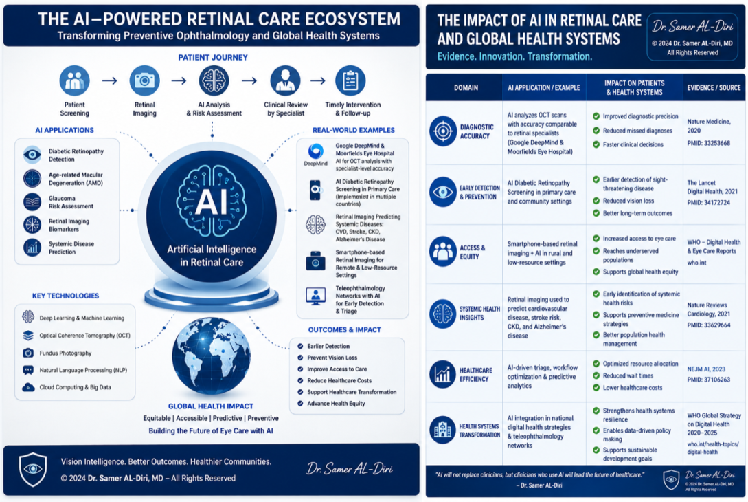
Artificial intelligence can support retinal screening, OCT interpretation and service planning, but clinical value depends on far more than algorithmic accuracy. Intended use, local validation, referral capacity, governance and continuous monitoring determine whether an AI tool improves care in practice.
Dr Samer Al-Diri
Ophthalmology and retinal care
Healthcare strategy and public health
**Key takeaways
Ophthalmology is data-rich**
Fundus photography and OCT create structured digital images that are well suited to machine-learning analysis.
**Regulated autonomy is narrow**
Autonomous diabetic-retinopathy screening shows that AI can perform a defined task, but it does not replace comprehensive eye examination.
**OCT evidence is promising**
Research systems have produced strong referral recommendations from OCT scans, although external and real-world validation remain essential.
**Foundation models broaden capability**
Large pretrained retinal models may reduce dependence on labelled data and support multiple downstream tasks, but generalisability cannot be assumed.
**Access requires a complete pathway**
Screening has little value without image-quality control, referral, treatment capacity, patient follow-up and accountability for missed disease.
**Governance is a clinical function**
Bias, privacy, cybersecurity, human oversight and post-deployment monitoring must be designed into the operating model.
**Why retinal imaging has become a leading field for clinical AI**
Ophthalmology combines high-volume digital imaging with clinically meaningful patterns that can be learned from data. Fundus photographs, OCT volumes, angiography and visual-field tests create opportunities for image classification, segmentation, triage and risk estimation.
The strongest use cases are not simply those with impressive technical performance. They are those in which the clinical question is clearly defined, the patient population is appropriate, the output leads to an agreed action and the health system can safely manage both positive and ungradable results.
**Strategic point:** an algorithm is only one component of a clinical service. The full intervention includes the device, staff, workflow, data infrastructure, referral rules, patient communication and access to treatment.
**Autonomous diabetic-retinopathy screening: an important but bounded model**
In 2018, the US Food and Drug Administration granted De Novo classification to IDx-DR, a device intended to detect more-than-mild diabetic retinopathy in a specified population of adults with diabetes. The associated pivotal trial enrolled 900 participants in primary-care settings.
This was a major regulatory milestone because the system could return a screening result without an eye-care specialist interpreting each image. Its intended use was nevertheless specific. It did not diagnose every retinal condition, screen for glaucoma or replace a full ophthalmic assessment. The FDA documentation also required clear action when disease was detected or when the system could not produce a result.
That distinction matters. Safe autonomy is not unrestricted automation; it is a tightly defined task supported by eligibility criteria, quality controls, referral pathways and professional accountability.
**OCT decision support and referral**
A landmark 2018 study developed a deep-learning system using 14,884 three-dimensional OCT scans from a clinically heterogeneous referral population. In the study setting, the system generated referral recommendations that reached or exceeded expert performance across several sight-threatening retinal diseases.
The study demonstrated the potential of OCT-based decision support, but high performance in a research evaluation is not the same as proven benefit across all clinics, devices and populations. Deployment still requires assessment of image quality, disease prevalence, calibration, false-negative risk, device compatibility and the consequences of errors.
**What health services should test before adoption**
• Whether the intended population matches the population used for development and validation.
• Performance across different devices, sites, ethnic groups and disease severities.
• How ungradable images, incidental findings and uncertain outputs are handled.
• Whether the service has sufficient referral and treatment capacity.
• Whether the tool improves patient outcomes, timeliness or equity—not only diagnostic accuracy.
**Foundation models and multimodal ophthalmic AI**
Retinal foundation models represent a shift from building a separate model for every narrow task toward pretraining on large image collections and adapting the resulting model to multiple applications. RETFound, reported in 2023, was pretrained on 1.6 million unlabelled retinal images and then adapted to ocular and systemic-disease tasks.
This approach may improve label efficiency and broaden reuse, especially where expertly annotated data are scarce. Multimodal systems may also combine fundus images, OCT, clinical history and other data. However, scale does not remove the need for clinical validation. A model that performs well in one dataset may fail when imaging protocols, equipment, case mix or care pathways change.
**Evidence boundary:** foundation models are an important research and development platform. They should not be described as universally generalisable or clinically interchangeable without task-specific and site-specific evidence.
**The retina as a source of systemic-health signals**
Deep-learning research has shown that retinal photographs contain signals associated with age, smoking status, blood pressure and other cardiovascular risk factors. This finding supports the scientific value of the retina as a non-invasive window into vascular and systemic health.
It does not mean that a retinal image can currently replace established cardiovascular assessment or laboratory testing. These models require prospective validation, calibration and evidence that their use improves decisions and outcomes. The responsible interpretation is that retinal imaging may contribute additional risk information—not that it provides a universal systemic diagnosis.
**Teleophthalmology and public-health value**
AI-assisted image assessment can help move parts of retinal screening closer to primary care and underserved communities. Portable cameras and remote review may reduce travel and make specialist capacity more targeted. The public-health opportunity is substantial: the World Health Organization estimates that at least 2.2 billion people have near or distance vision impairment, and at least 1 billion cases could have been prevented or have yet to be addressed.
Technology alone will not close this gap. A credible programme needs population eligibility rules, community engagement, trained operators, image-quality assurance, rapid referral, treatment access and monitoring of who is lost between screening and care.
**Measures that matter at system level**
• Coverage among the eligible population.
• Ungradable-image rate and repeat-imaging rate.
• Time from screening to specialist assessment.
• Referral completion and treatment completion.
• Performance and access across population groups.
• Patient-reported experience and accessibility.
**Generative AI: useful assistance, different risks**
Generative AI may assist with draft reports, patient information, documentation and workflow coordination. These applications are different from a validated diagnostic device. Generated text can be incomplete, inaccurate or overconfident, and may reproduce bias or expose sensitive information if governance is weak.
The World Health Organization’s guidance on large multimodal models in health emphasises transparency, human responsibility, rigorous evaluation, data protection and appropriate regulation. In ophthalmology, any generated clinical content should remain traceable to source data, reviewed by an accountable professional and prevented from silently changing the diagnostic record.
**A practical governance model for implementation
Governance question** **Required operational response**
What exactly is the tool authorised and intended to do? Define the population, clinical task, exclusions, output and action threshold.
Does it work safely here? Conduct local validation and workflow testing before routine deployment.
Who remains accountable? Name the responsible clinical and operational owners and specify escalation rules.
What happens when the system is uncertain or fails? Create pathways for ungradable images, no-result outputs, downtime and suspected errors.
Could performance differ across groups? Monitor calibration, false-negative rates and access by relevant demographic and clinical groups.
How will change be controlled? Review software updates, device changes, model drift, cybersecurity and post-market evidence.
**Implementation sequence for healthcare leaders
1. Start with the service problem.** Define the unmet need, baseline performance and intended patient benefit.
**2. Specify the use case.** Set the population, setting, exclusions, decision threshold and human role.
**3. Assess evidence and regulation.** Review authorisation, external validation, limitations and relevance to the local service.
**4. Test the complete pathway.** Include imaging, data transfer, result communication, referral, treatment and downtime arrangements.
**5. Deploy in stages.** Use controlled implementation with staff training, incident reporting and clear stop criteria.
**6. Monitor value continuously.** Track safety, equity, outcomes, workflow effects, cost and patient experience.
**Frequently asked questions
Will AI replace ophthalmologists?**
No. Current systems perform defined tasks. Ophthalmologists remain responsible for clinical context, complex diagnosis, shared decisions, treatment and accountability.
**Is autonomous AI already used in eye care?**
Yes, regulated autonomous systems exist for specific diabetic-retinopathy screening indications. Their authorisation does not extend to comprehensive eye examination or every retinal disease.
**Can AI interpret OCT scans?**
Research systems have shown strong performance in OCT classification, segmentation and referral support. Safe routine use depends on the authorised purpose, external validation and the local clinical pathway.
**What is the main implementation risk?**
The greatest risk is treating algorithmic performance as if it were a complete service. Weak referral capacity, poor data quality, unclear accountability or inequitable access can undermine an otherwise capable tool.
**Conclusion**
AI-powered retinal imaging has moved beyond proof of concept in selected applications, particularly diabetic-retinopathy screening, while OCT decision support and retinal foundation models continue to advance. The strategic question is no longer whether AI can analyse images. It is whether a health system can convert that capability into safer, more timely and more equitable care.
The strongest programmes will combine evidence, regulation, clinical leadership, public-health design and continuous governance. That is how technical performance becomes dependable patient value.
**Declarations
Conflict of interest:** The author declares no conflict of interest related to this article.
**Clinical disclaimer:** This article is for professional education and general information. It does not replace individual clinical assessment, medical advice, device labelling or local regulatory requirements.
**Evidence note:** The article distinguishes established regulatory or research findings from emerging applications and strategic interpretation. Selected evidence and authoritative guidance were reviewed through 22 August 2026.
**References and authoritative sources**
1. World Health Organization. <u>[World](who.int/publications-detail-redirect/world-report-on-vision)[report](who.int/publications-detail-redirect/world-report-on-vision)[on](who.int/publications-detail-redirect/world-report-on-vision)[vision](who.int/publications-detail-redirect/world-report-on-vision)</u>. 2019.
2. Abràmoff MD, Lavin PT, Birch M, Shah N, Folk JC. <u>[Pivotal](doi.org/10.1038/s41746-018-0040-6)[](doi.org/10.1038/s41746-018-0040-6)[trial](doi.org/10.1038/s41746-018-0040-6)[of an](doi.org/10.1038/s41746-018-0040-6)[autonomous](doi.org/10.1038/s41746-018-0040-6)[](doi.org/10.1038/s41746-018-0040-6)[AI-based](doi.org/10.1038/s41746-018-0040-6)[](doi.org/10.1038/s41746-018-0040-6)[diagnostic](doi.org/10.1038/s41746-018-0040-6)[](doi.org/10.1038/s41746-018-0040-6)[system](doi.org/10.1038/s41746-018-0040-6)[for](doi.org/10.1038/s41746-018-0040-6)[detection](doi.org/10.1038/s41746-018-0040-6)[of](doi.org/10.1038/s41746-018-0040-6)[diabetic](doi.org/10.1038/s41746-018-0040-6)[](doi.org/10.1038/s41746-018-0040-6)[retinopathy](doi.org/10.1038/s41746-018-0040-6)[in](doi.org/10.1038/s41746-018-0040-6)[primary](doi.org/10.1038/s41746-018-0040-6)[](doi.org/10.1038/s41746-018-0040-6)[care](doi.org/10.1038/s41746-018-0040-6)[](doi.org/10.1038/s41746-018-0040-6)[offices](doi.org/10.1038/s41746-018-0040-6)</u>. npj Digital Medicine. 2018;1:39.
3. US Food and Drug Administration. <u>[De Novo](accessdata.fda.gov/cdrh_docs/reviews/DEN180001.pdf)[classification](accessdata.fda.gov/cdrh_docs/reviews/DEN180001.pdf)[](accessdata.fda.gov/cdrh_docs/reviews/DEN180001.pdf)[request](accessdata.fda.gov/cdrh_docs/reviews/DEN180001.pdf)[for](accessdata.fda.gov/cdrh_docs/reviews/DEN180001.pdf)[IDx-DR](accessdata.fda.gov/cdrh_docs/reviews/DEN180001.pdf)[:](accessdata.fda.gov/cdrh_docs/reviews/DEN180001.pdf)[decision](accessdata.fda.gov/cdrh_docs/reviews/DEN180001.pdf)[](accessdata.fda.gov/cdrh_docs/reviews/DEN180001.pdf)[summary](accessdata.fda.gov/cdrh_docs/reviews/DEN180001.pdf)[(DEN180001)](accessdata.fda.gov/cdrh_docs/reviews/DEN180001.pdf)</u>. 2018.
4. De Fauw J, Ledsam JR, Romera-Paredes B, et al. <u>[Clinically](doi.org/10.1038/s41591-018-0107-6)[](doi.org/10.1038/s41591-018-0107-6)[applicable](doi.org/10.1038/s41591-018-0107-6)[](doi.org/10.1038/s41591-018-0107-6)[deep](doi.org/10.1038/s41591-018-0107-6)[](doi.org/10.1038/s41591-018-0107-6)[learning](doi.org/10.1038/s41591-018-0107-6)[for](doi.org/10.1038/s41591-018-0107-6)[diagnosis](doi.org/10.1038/s41591-018-0107-6)[and](doi.org/10.1038/s41591-018-0107-6)[referral](doi.org/10.1038/s41591-018-0107-6)[in](doi.org/10.1038/s41591-018-0107-6)[retinal](doi.org/10.1038/s41591-018-0107-6)[](doi.org/10.1038/s41591-018-0107-6)[disease](doi.org/10.1038/s41591-018-0107-6)</u>. Nature Medicine. 2018;24:1342–1350.
5. Zhou Y, Chia MA, Wagner SK, et al. <u>[A](doi.org/10.1038/s41586-023-06555-x)[foundation](doi.org/10.1038/s41586-023-06555-x)[](doi.org/10.1038/s41586-023-06555-x)[model](doi.org/10.1038/s41586-023-06555-x)[for](doi.org/10.1038/s41586-023-06555-x)[generalizable](doi.org/10.1038/s41586-023-06555-x)[](doi.org/10.1038/s41586-023-06555-x)[disease](doi.org/10.1038/s41586-023-06555-x)[](doi.org/10.1038/s41586-023-06555-x)[detection](doi.org/10.1038/s41586-023-06555-x)[from](doi.org/10.1038/s41586-023-06555-x)[retinal](doi.org/10.1038/s41586-023-06555-x)[](doi.org/10.1038/s41586-023-06555-x)[images](doi.org/10.1038/s41586-023-06555-x)</u>. Nature. 2023;622:156–163.
6. Poplin R, Varadarajan AV, Blumer K, et al. <u>[Prediction](doi.org/10.1038/s41551-018-0195-0)[of](doi.org/10.1038/s41551-018-0195-0)[cardiovascular](doi.org/10.1038/s41551-018-0195-0)[](doi.org/10.1038/s41551-018-0195-0)[risk](doi.org/10.1038/s41551-018-0195-0)[](doi.org/10.1038/s41551-018-0195-0)[factors](doi.org/10.1038/s41551-018-0195-0)[from](doi.org/10.1038/s41551-018-0195-0)[retinal](doi.org/10.1038/s41551-018-0195-0)[](doi.org/10.1038/s41551-018-0195-0)[fundus](doi.org/10.1038/s41551-018-0195-0)[](doi.org/10.1038/s41551-018-0195-0)[photographs](doi.org/10.1038/s41551-018-0195-0)[](doi.org/10.1038/s41551-018-0195-0)[via](doi.org/10.1038/s41551-018-0195-0)[](doi.org/10.1038/s41551-018-0195-0)[deep](doi.org/10.1038/s41551-018-0195-0)[](doi.org/10.1038/s41551-018-0195-0)[learning](doi.org/10.1038/s41551-018-0195-0)</u>. Nature Biomedical Engineering. 2018;2:158–164.
7. World Health Organization. <u>[Ethics](who.int/publications/i/item/9789240029200)[and](who.int/publications/i/item/9789240029200)[governance](who.int/publications/i/item/9789240029200)[of](who.int/publications/i/item/9789240029200)[artificial](who.int/publications/i/item/9789240029200)[](who.int/publications/i/item/9789240029200)[intelligence](who.int/publications/i/item/9789240029200)[for](who.int/publications/i/item/9789240029200)[health](who.int/publications/i/item/9789240029200)</u>. 2021.
8. World Health Organization. <u>[Ethics](who.int/publications/i/item/9789240084759)[and](who.int/publications/i/item/9789240084759)[governance](who.int/publications/i/item/9789240084759)[of](who.int/publications/i/item/9789240084759)[artificial](who.int/publications/i/item/9789240084759)[](who.int/publications/i/item/9789240084759)[intelligence](who.int/publications/i/item/9789240084759)[for](who.int/publications/i/item/9789240084759)[health](who.int/publications/i/item/9789240084759)[:](who.int/publications/i/item/9789240084759)[guidance](who.int/publications/i/item/9789240084759)[on](who.int/publications/i/item/9789240084759)[large](who.int/publications/i/item/9789240084759)[](who.int/publications/i/item/9789240084759)[multi-modal](who.int/publications/i/item/9789240084759)[](who.int/publications/i/item/9789240084759)[models](who.int/publications/i/item/9789240084759)</u>. 2024.
9. US Food and Drug Administration. <u>[Artificial](fda.gov/medical-devices/software-medical-device-samd/artificial-intelligence-enabled-medical-devices)[](fda.gov/medical-devices/software-medical-device-samd/artificial-intelligence-enabled-medical-devices)[Intelligence-Enabled](fda.gov/medical-devices/software-medical-device-samd/artificial-intelligence-enabled-medical-devices)[Medical Devices](fda.gov/medical-devices/software-medical-device-samd/artificial-intelligence-enabled-medical-devices)</u>. Accessed 22 August 2026.
**About Dr Samer Al-Diri**
Dr. Samer Al-Diri, MD, MSc, MPH, is a UK-trained ophthalmologist, public-health professional and strategic healthcare consultant. His work spans healthcare management, health-system transformation, clinical governance, prevention, responsible digital health and eye health.
<u>[Website](drsameraldiri.com/)</u>
<u>[LinkedIn](linkedin.com/in/dr-samer-al-diri/)</u>
<u>[ORCID](orcid.org/0009-0004-1908-0714)</u>
<u>[ResearchGate](researchgate.net/profile/Samer-Al-Diri)</u>
<u>[Google](scholar.google.com/citations?user=AeyXd5MAAAAJ)[Scholar](scholar.google.com/citations?user=AeyXd5MAAAAJ)</u>